Full Text
Introduction
Acute disseminated encephalomyelitis (ADEM) is a rare, acute, immune-mediated inflammatory demyelinating disorder of the central nervous system (CNS). It is characterised clinically by acute encephalopathy accompanied by multifocal neurological manifestations and radiologically by multifocal inflammatory abnormalities within the brain and, in some cases, the spinal cord and optic pathways. ADEM is most frequently recognised in children and adolescents, but adult-onset disease is well documented and may present with considerable diagnostic difficulty because its clinical manifestations overlap with infectious encephalitis, autoimmune encephalitis, multiple sclerosis, neuromyelitis optica spectrum disorder, and other inflammatory CNS disorders [1,2].
The disorder is generally monophasic and may occur after a preceding infection or, less commonly, following immunisation. Historically, ADEM has been reported in association with several infectious illnesses and vaccines. Vaccination-associated ADEM remains uncommon, and a temporal relationship between vaccination and neurological symptoms should be interpreted cautiously because temporal association alone cannot establish causality [1,3].
The pathogenesis of ADEM is incompletely understood. Current evidence supports an immune-mediated process in which an environmental trigger, such as infection or immunisation, may initiate an inflammatory response directed against CNS myelin in susceptible individuals. Molecular mimicry and other dysregulated immune mechanisms have been proposed, although no single mechanism explains all cases [3]. The absence of a disease-specific biomarker further complicates diagnosis, particularly in adults, and diagnosis therefore depends on the clinical presentation, neuroimaging findings, exclusion of alternative diagnoses, and clinical course [2-4].
Clinical manifestations are heterogeneous. Fever, headache, nausea, vomiting, fatigue, altered behaviour, seizures, sensory symptoms, motor deficits, ataxia, dysarthria, visual symptoms, and other focal neurological abnormalities may occur. Encephalopathy is an important clinical feature and may range from irritability and confusion to marked alteration of consciousness [2,5]. MRI is the preferred imaging modality for evaluating suspected ADEM, although imaging findings may vary with disease stage and may occasionally be initially normal. CSF analysis may demonstrate lymphocytic pleocytosis and elevated protein, but these findings are nonspecific and mainly assist in evaluating alternative infectious or inflammatory diagnoses [5,6].
Treatment is directed toward suppressing the acute inflammatory process. High-dose intravenous corticosteroids are generally used as first-line therapy, followed by a tapering course of oral corticosteroids. Intravenous immunoglobulin or plasma exchange may be considered in patients who fail to respond adequately to corticosteroids or who have severe or refractory disease [7]. Because infectious encephalitis and other potentially treatable conditions can mimic ADEM, appropriate microbiological and molecular investigations should be undertaken before or concurrently with immunosuppressive treatment when clinically indicated.
Human papillomavirus (HPV) vaccination is an important preventive intervention against HPV infection and HPV-associated malignancies. Neurological adverse events following HPV vaccination are uncommon, and reports of ADEM following HPV vaccination are rare [3]. Published case reports have described ADEM following different HPV vaccine formulations, but the rarity of the event makes individual cases important for clinical awareness while not establishing a causal relationship. We report an adult woman who developed an acute encephalopathic and multifocal neurological syndrome shortly after receiving Gardasil 9 and who improved rapidly following corticosteroid therapy.
The objective of this case report is to describe the clinical presentation, diagnostic evaluation, treatment, and short-term outcome of suspected post-vaccination ADEM in an adult patient.
Case presentation
A 26-year-old woman with no known comorbidities or previous neurological illness presented with fever and headache of 6 days' duration. The symptoms reportedly began approximately 6 hours after administration of the Gardasil 9 HPV vaccine. She was initially evaluated at another hospital, where routine blood investigations were reported to be within normal limits. Non-contrast MRI of the brain and magnetic resonance venography were also reported as normal. Despite symptomatic treatment, her symptoms persisted, prompting presentation to our hospital for further evaluation. Informed consent was obtained from the patient for publication of the case report and accompanying clinical information/images.
At admission, she was conscious and coherent, with a temperature of 100.5°F; the remaining vital signs were within normal limits. There were no focal neurological deficits or clinical signs of meningeal irritation at the time of initial examination. She was initially treated with intravenous ceftriaxone 2 g because an infectious etiology remained an important consideration.
During hospitalisation, she developed sudden worsening of headache, numbness of the tongue, and inability to speak, followed by irritability and altered sensorium the following day. In view of the acute neurological deterioration, an extensive diagnostic evaluation was undertaken, including CSF analysis, contrast-enhanced MRI of the brain, and whole-body 18F-FDG PET/CT.
Complete blood count, urine examination, C-reactive protein, thyroid profile, liver and kidney function tests, and antinuclear antibody testing were unremarkable. Viral screening, investigations for tropical infections, blood cultures, and procalcitonin were negative. Ultrasonography of the abdomen and 2D echocardiography were normal.
CSF examination showed 576 white blood cells/µL, with lymphocytes accounting for 85% of the cells, glucose of 44 mg/dL, and protein of 193.5 mg/dL. Acid-fast bacillus testing, Mycobacterium tuberculosis GeneXpert, and a meningoencephalitis PCR panel for common bacterial and viral pathogens were negative (Table 1). CSF culture was also negative. Testing for myelin oligodendrocyte glycoprotein (MOG) antibodies and aquaporin-4 antibodies was negative.
Table 1: Meningoencephalitis Biofire panel report.
|
Parameter
|
Result
|
Normal
|
|
Investigation
|
Meningoencephalitis panel
|
null
|
|
Sample type
|
CSF
|
null
|
|
Method
|
Multiplex PCR
|
null
|
|
Result
|
Not detected
|
null
|
|
Escherichia coli K1
|
Not detected
|
null
|
|
Streptococcus pneumoniae
|
Not detected
|
null
|
|
Streptococcus agalactiae
|
Not detected
|
null
|
|
Listeria monocytogenes
|
Not detected
|
null
|
|
Neisseria meningitidis
|
Not detected
|
null
|
|
Haemophilus influenzae
|
Not detected
|
null
|
|
Cryptococcus neoformans/GATTI
|
Not detected
|
null
|
|
Cytomegalovirus
|
Not detected
|
null
|
|
Entero virus
|
Not detected
|
null
|
|
Herpes simplex - 1
|
Not detected
|
null
|
|
Herpes simplex - 2
|
Not detected
|
null
|
|
Human herpesvirus 6
|
Not detected
|
null
|
|
Human parechovirus
|
Not detected
|
null
|
|
Varicella zoster virus
|
Not detected
|
null
|
Contrast-enhanced MRI of the brain demonstrated diffuse leptomeningeal enhancement along the frontal, parietal, and temporal convexities, cerebellar folia, and pial surface of the brainstem (Figure 1). 18F-FDG PET/CT of the brain demonstrated intensely hypermetabolic foci involving the vermis and right temporal lobe, with moderately intense hypermetabolism involving the left insular cortex, anterior cingulate gyrus, and left cerebellum. Hypometabolic areas were also noted in the bilateral posterior parietal, left temporal, bilateral parieto-occipital regions, and bilateral thalami. The PET-CT findings were interpreted as suggestive of an autoimmune inflammatory encephalitic process (Figure 2).
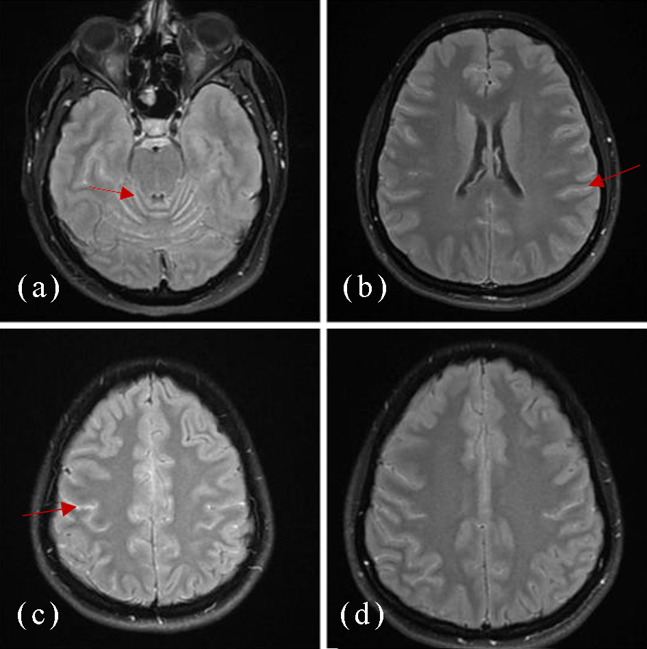
Figure 1: Post-contrast FLAIR images showing diffuse leptomeningeal enhancement.
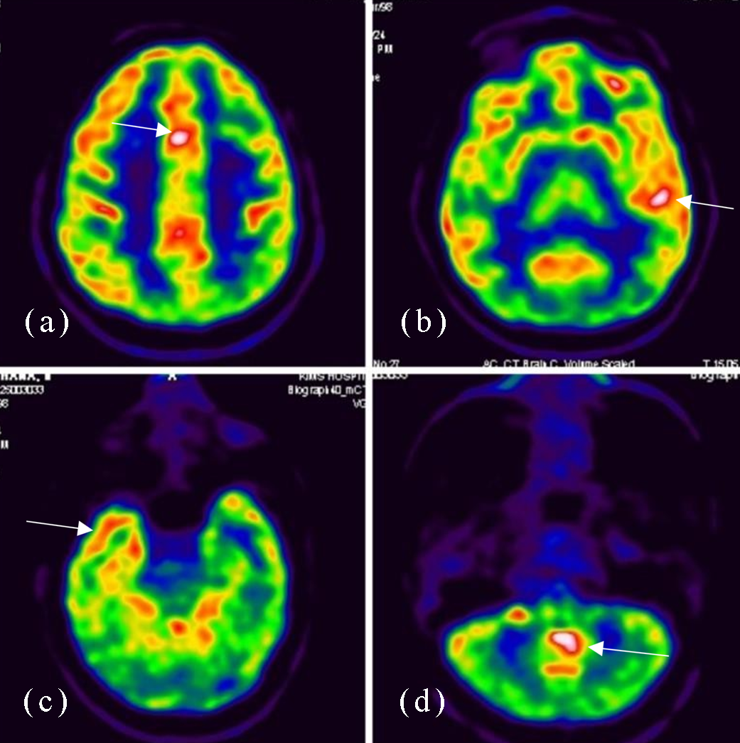
Figure 2: FDG PET-CT brain images showing (clockwise from the upper-left image) hypermetabolism in the anterior cingulate gyrus, left insular cortex, vermis, and right temporal region.
Given the acute encephalopathic presentation, multifocal neurological manifestations, inflammatory CSF findings, imaging abnormalities, negative infectious evaluation, negative MOG and aquaporin-4 antibody testing, and the temporal relationship to HPV vaccination, we considered suspected post-vaccination ADEM. The temporal relationship was regarded as supportive clinical context rather than proof of vaccine causality.
The patient received intravenous methylprednisolone at a dose of 1 g daily for 5 days, together with intravenous ceftriaxone and levetiracetam. Her neurological symptoms resolved rapidly following corticosteroid therapy. She was discharged in a clinically improved condition on an oral tapering regimen of corticosteroids. No neurological relapse was documented during the follow-up described in the submitted case material.
Discussion
ADEM is an acute inflammatory demyelinating CNS disorder that typically presents with a rapid onset of encephalopathy and multifocal neurological deficits. Although it is predominantly a pediatric disorder, adult patients may develop ADEM. They can present with clinical features that overlap with infectious encephalitis, autoimmune encephalitis, multiple sclerosis, and other inflammatory disorders [1,2]. The present case illustrates the diagnostic challenge of ADEM in an adult who initially presented with fever and headache and subsequently developed focal neurological symptoms and altered sensorium.
The temporal relationship between vaccination and symptom onset was striking in this patient, with fever and headache beginning approximately 6 hours after administration of Gardasil 9. However, the close temporal association should not be interpreted as definitive evidence of causality. ADEM has been reported after immunisation, but the underlying mechanisms remain incompletely understood, and coincidental onset of an inflammatory CNS disorder after vaccination is possible [1,3]. Previous reports have described ADEM following HPV vaccination, demonstrating that such an association has been observed, although it remains rare.
The diagnostic evaluation was important because infection is a major differential diagnosis in a patient with fever, headache, CSF pleocytosis, and altered sensorium. The patient underwent extensive infectious evaluation, including a meningoencephalitis PCR panel, CSF culture, acid-fast bacillus testing, Mycobacterium tuberculosis GeneXpert, blood cultures, viral screening, and assessment of inflammatory markers. These investigations did not identify an infectious cause. MOG and aquaporin-4 antibodies were also negative, reducing support for MOG-associated disease and neuromyelitis optica spectrum disorder, although antibody negativity alone does not exclude all inflammatory demyelinating conditions.
MRI is an important component of the evaluation of suspected ADEM. Typical ADEM lesions are multifocal and may have poorly defined margins, although imaging findings can vary depending on disease stage and patient age [2,5]. In the present patient, the initial MRI was reported as normal, whereas subsequent contrast-enhanced MRI demonstrated diffuse leptomeningeal enhancement. This evolution emphasises that an initially unrevealing MRI does not necessarily exclude an evolving inflammatory CNS disorder. Nevertheless, leptomeningeal enhancement is not specific for ADEM and can be seen in several infectious, inflammatory, and neoplastic conditions; therefore, the imaging findings must be interpreted together with the clinical course and exclusion of alternative diagnoses.
The CSF profile showed marked lymphocytic pleocytosis and elevated protein. Although CSF abnormalities may occur in ADEM, there is no specific CSF biomarker that establishes the diagnosis [2,5,6]. The substantial lymphocytic pleocytosis in this case further justified the extensive evaluation for infectious meningoencephalitis before immunosuppressive treatment. The absence of identifiable infectious pathogens, together with the clinical and imaging findings and subsequent response to corticosteroid therapy, supported an immune-mediated inflammatory diagnosis.
High-dose intravenous corticosteroids remain the principal first-line treatment for ADEM. The patient received intravenous methylprednisolone 1 g daily for 5 days and demonstrated rapid clinical improvement, followed by discharge on an oral steroid taper. Published guidance and reviews support high-dose corticosteroids as initial therapy, with intravenous immunoglobulin or plasma exchange considered for patients with inadequate response or severe refractory disease [7]. The favourable response in this case is consistent with the expected treatment response of an acute inflammatory demyelinating syndrome, although treatment response alone cannot establish the diagnosis.
This case has several limitations. First, a single case cannot establish a causal relationship between HPV vaccination and ADEM. Second, the imaging findings were not entirely typical of classic ADEM, and the initial MRI was normal. Third, although an extensive infectious evaluation was negative, no single investigation can completely exclude every possible infectious or inflammatory mimic. Finally, the follow-up information available in the submitted case material is limited, restricting assessment of long-term neurological outcome or recurrence.
Despite these limitations, the case is clinically relevant because it demonstrates the importance of maintaining a broad differential diagnosis in an adult with acute encephalopathy and multifocal neurological manifestations after immunisation. Early recognition, exclusion of infectious causes, appropriate neuroimaging, CSF evaluation, and timely immunotherapy may contribute to favourable clinical recovery.
Conclusion
Suspected ADEM should be considered in adults who develop acute encephalopathy and multifocal neurological manifestations following an infection or immunisation. In patients with a temporal association with HPV vaccination, clinicians should undertake appropriate investigations to exclude infectious, autoimmune, and other neurological causes before attributing the presentation to vaccination. In this case, extensive evaluation did not identify an infectious or alternative autoimmune cause, and the patient improved rapidly following high-dose intravenous corticosteroid therapy. The rarity of reported post-HPV-vaccination ADEM should not discourage recommended HPV immunisation, but awareness of this uncommon inflammatory neurological presentation may facilitate early diagnosis and treatment.
Acknowledgements
Dr. Praveen Yada, neurologist and the Departments of Laboratory, Radiology, and Nuclear Medicine.
Conflict of interest
The authors declare that they have no conflict of interest.
References
[1] Pohl D, Alper G, Van Haren K, Kornberg AJ, Lucchinetti CF, et al. Acute disseminated encephalomyelitis: updates on an inflammatory CNS syndrome. Neurology. 2016; 87: 38–45.
[2] Paolilo RB, Deiva K, Neuteboom R, Rostásy K, Lim M. Acute disseminated encephalomyelitis: current perspectives. Children (Basel). 2020; 7: 210.
[3] Menge T, Kieseier BC, Nessler S, Hemmer B, Hartung HP, et al. Acute disseminated encephalomyelitis: an acute hit against the brain. Curr Opin Neurol. 2007; 20:247–254.
[4] Lin C, Jeng J, Hsieh S. Acute disseminated encephalomyelitis: a follow-up study in Taiwan. J Neurol Neurosurg Psychiatry. 2007; 78:162–167.
[5] Paolilo RB, Deiva K, Neuteboom R, Rostásy K, Lim M. Acute disseminated encephalomyelitis: current perspectives. Children (Basel). 2020; 7:210.
[6] Anilkumar AC, Foris LA, Tadi P. Acute disseminated encephalomyelitis. In: StatPearls [Internet]. Treasure Island (FL): StatPearls Publishing; 2025.
[7] Pohl D, Tenembaum S. Treatment of acute disseminated encephalomyelitis. Curr Treat Options Neurol. 2012; 14:264–275.
[8] Bhatt P, Bray L, Raju S, Dapaah-Siakwan F, Patel A, et al. Temporal trends of pediatric hospitalizations with acute disseminated encephalomyelitis in the United States: an analysis from 2006 to 2014 using National Inpatient Sample. J Pediatr. 2019; 206:26–32.