Full Text
Introduction
Malaria has a broad clinical spectrum, ranging from asymptomatic parasitaemia to severe, life-threatening disease. Plasmodium falciparum is particularly associated with severe multisystem complications, including cerebral malaria, acute kidney injury, severe anaemia, acute respiratory distress syndrome, metabolic disturbances, hepatic dysfunction, and circulatory shock [1–3].
Cardiovascular involvement in malaria is less frequently recognized than other complications. Reported manifestations include myocarditis, pericarditis, arrhythmias, conduction abnormalities, myocardial ischaemia, and heart failure [4,5]. Cardiovascular manifestations may result from systemic inflammation, endothelial dysfunction, sequestration of parasitized erythrocytes, tissue hypoxia, and metabolic disturbances. In addition, some antimalarial agents may produce cardiovascular adverse effects, further complicating clinical assessment [5].
Patients with pre-existing structural heart disease may be particularly vulnerable to haemodynamic deterioration during acute systemic infection. We report a case of acute cardiac decompensation in a patient with severe rheumatic mitral stenosis and tricuspid regurgitation during P. falciparum malaria in a resource-limited field hospital in South Sudan. The case highlights the diagnostic and therapeutic challenges associated with acute cardiovascular deterioration in a patient with significant underlying valvular heart disease and concomitant malaria.
Case presentation
A 46-year-old woman presented to our field hospital with a 5-day history of fever associated with chills, rigors, body ache, and headache. She had a history of rheumatic heart disease diagnosed in 2011 and had previously undergone balloon mitral valvotomy. On admission, she was haemodynamically stable, and her general examination did not reveal any significant abnormality. Written informed consent obtained from the patient for publication of this case report and the accompanying images.
A malaria rapid diagnostic test was positive for P. falciparum (Figure 1a). The diagnosis was confirmed by thin peripheral blood smear, which demonstrated P. falciparum schizonts within infected erythrocytes (Figure 1b).
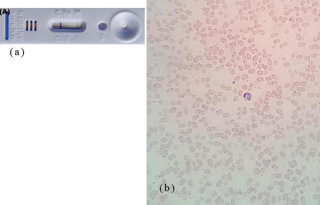
Figure 1: Diagnosis of Plasmodium falciparum malaria. (A) Malaria rapid diagnostic test positive for P. falciparum. (B) Thin peripheral blood smear demonstrating P. falciparum schizonts within infected erythrocytes.
Clinical deterioration
The patient was initiated on artemisinin-based antimalarial therapy and antipyretics. Approximately 16 hours after admission, she developed acute-onset orthopnoea, corresponding to New York Heart Association functional class IV symptoms, together with cough producing pink frothy sputum. She also reported chest pain, palpitations, and syncope. Her heart rate was 110 beats/min, blood pressure was 90/40 mmHg, oxygen saturation was 80% on room air and improved to 90% with oxygen at 6 L/min through a face mask. Her respiratory rate was 30 breaths/min.
Cardiovascular examination revealed a grade III mid-diastolic rumbling murmur with presystolic accentuation, best heard at the cardiac apex in the mitral area. Bilateral coarse crackles were present throughout both lung fields.
Investigations
Laboratory investigations showed leucocytosis (11,800/mm³), markedly elevated C-reactive protein (CRP), prolonged prothrombin time (16.7 seconds; INR, 1.26), elevated D-dimer (3740 µg/L), and hyponatraemia (serum sodium, 124 mEq/L). Platelet count and serum potassium were within normal limits. Cardiac enzyme levels were normal, and NT-proBNP was 71.6 pg/mL.
Posteroanterior chest radiography demonstrated cardiomegaly with a cardiothoracic ratio of approximately 0.51 and radiographic features of left atrial enlargement, including an increased carinal angle of 106° and right retrocardiac double density (Figure 2). No definite radiographic evidence of pulmonary oedema was identified.
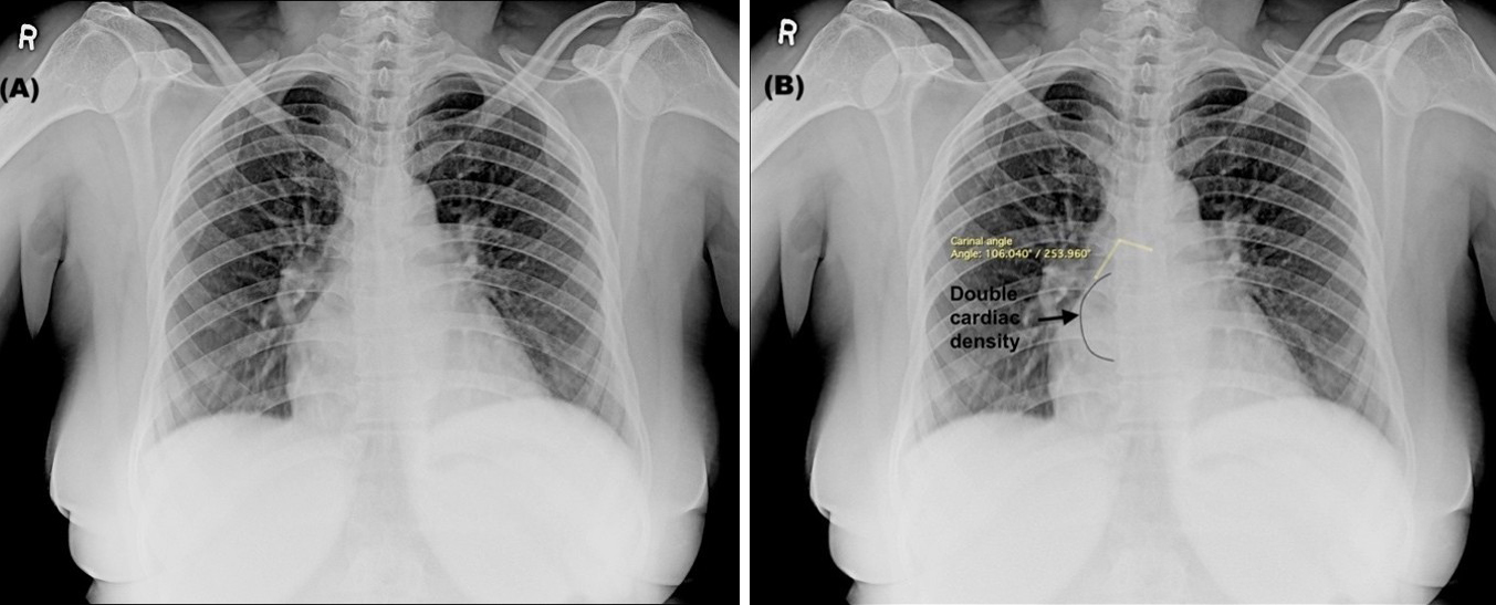
Figure 2: Posteroanterior chest radiograph (a) Unannotated image, (b) Annotated image demonstrating cardiomegaly and features of left atrial enlargement. No definite pulmonary oedema is seen.
Electrocardiography demonstrated sinus tachycardia with P pulmonale (Figure 3). Urgent two-dimensional echocardiography demonstrated severe rheumatic mitral stenosis, severe tricuspid regurgitation, and severe pulmonary arterial hypertension, with a peak systolic tricuspid regurgitation pressure gradient of 71 mmHg. The left atrium was moderately enlarged, measuring 5 cm, while the right atrium and right ventricle were dilated. Left ventricular systolic function was preserved, with an ejection fraction of 70%.
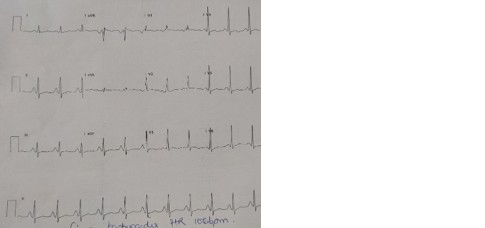
Figure 3: Electrocardiogram demonstrating sinus tachycardia and P pulmonale.
Treatment and outcome
The patient was managed with antimalarial therapy, intramuscular benzathine penicillin (1.2 million units monthly), intravenous dobutamine infusion (10 mg/h, titrated according to haemodynamic response), oral digoxin, furosemide, sildenafil, and rivaroxaban. She was maintained in a propped-up position and received supplemental oxygen.
Clinical improvement was observed within 12 hours of initiating treatment, and her vital parameters normalized within 36 hours. After 7 days of hospitalization, she was discharged in a clinically stable condition and referred to a higher centre for definitive management of her mitral and tricuspid valve disease.
Discussion
Malaria is a multisystem infectious disease with manifestations ranging from uncomplicated parasitaemia to severe, life-threatening illness. P. falciparum is particularly associated with severe disease because of its capacity for erythrocyte sequestration, endothelial interaction, and systemic inflammatory activation [1,2]. Cardiovascular involvement is less frequently recognized but encompasses a broad spectrum of abnormalities, including myocarditis, pericarditis, arrhythmias, conduction disturbances, myocardial ischaemia, and heart failure [4,5].
The cardiovascular manifestations of malaria are likely multifactorial. Proposed mechanisms include systemic inflammatory responses, endothelial dysfunction, sequestration of parasitized erythrocytes in the microcirculation, impaired tissue oxygenation, metabolic disturbances, and direct or indirect myocardial injury [5-9]. Cytokine-mediated myocardial dysfunction has also been proposed, with severe malaria associated with increased circulating concentrations of pro- and anti-inflammatory cytokines [8]. Anaemia, renal dysfunction, altered intravascular volume, and other systemic complications may further increase cardiovascular stress [10–12].
Our patient had significant pre-existing structural heart disease, including severe rheumatic mitral stenosis, severe tricuspid regurgitation, and severe pulmonary arterial hypertension. These abnormalities provide a plausible substrate for cardiac decompensation during an acute systemic infection. The temporal relationship between P. falciparum infection and the sudden development of orthopnoea, hypoxaemia, hypotension, and pulmonary congestion-like symptoms suggests that the acute infection may have acted as a precipitating stressor. However, the available findings do not establish direct myocardial injury due to malaria. Therefore, the presentation is more appropriately interpreted as acute cardiac decompensation occurring in the setting of falciparum malaria and severe underlying valvular heart disease.
The absence of elevated cardiac enzymes and the preserved left ventricular ejection fraction argue against an overt myocardial infarction or severe global left ventricular systolic dysfunction. The normal NT-proBNP concentration also makes interpretation of the acute heart failure syndrome less straightforward. Nevertheless, natriuretic peptide concentrations should be interpreted in the context of the clinical presentation and underlying valvular pathology rather than in isolation. The ECG showed sinus tachycardia and P pulmonale, findings compatible with the patient's haemodynamic burden and pulmonary hypertension but not specific for malaria-related myocardial involvement.
Cardiovascular abnormalities in malaria may be under-recognized because clinical manifestations can overlap with severe systemic infection and pre-existing cardiovascular disease. Gupta et al. emphasized the range of cardiovascular manifestations associated with malaria and the potential role of systematic cardiovascular assessment [7]. A nationwide Danish cohort study also reported an association between previous imported malaria, particularly P. falciparum infection, and subsequent risk of heart failure, although the study addressed long-term cardiovascular outcomes rather than acute cardiac decompensation [4].
Echocardiography was particularly valuable in this case because it demonstrated the severity of the underlying valvular disease and pulmonary hypertension. In a patient with malaria who develops unexplained dyspnoea, hypotension, hypoxaemia, chest discomfort, palpitations, or other cardiovascular symptoms, careful clinical assessment supplemented by ECG, cardiac biomarkers, and echocardiography may help identify clinically important cardiac involvement and distinguish it from other complications of severe malaria.
The management of this patient was challenging because definitive valvular intervention was not immediately available in the resource-limited field-hospital setting. Initial treatment therefore focused on antimalarial therapy, haemodynamic support, diuresis, oxygen supplementation, and management of the underlying cardiovascular condition. The patient's rapid clinical improvement following treatment supports the importance of early recognition and stabilization while arranging definitive cardiac evaluation.
This case has important limitations. It is a single-patient observation, and the temporal association between malaria and cardiac decompensation does not establish causality. The patient had severe pre-existing rheumatic valvular disease, which itself provides a strong explanation for susceptibility to acute haemodynamic deterioration. In addition, advanced cardiac investigations such as cardiac magnetic resonance imaging and invasive haemodynamic assessment were not available in the field setting. These limitations should be acknowledged when interpreting the case.
Overall, this case illustrates that P. falciparum malaria may precipitate clinically significant cardiovascular deterioration in patients with severe underlying structural heart disease. A high index of suspicion and early cardiovascular evaluation are warranted when patients with malaria develop acute cardiorespiratory symptoms.
Conclusion
P. falciparum malaria may precipitate acute cardiac decompensation in patients with severe underlying rheumatic valvular heart disease. In this case, severe mitral stenosis, tricuspid regurgitation, and pulmonary hypertension provided a substantial substrate for haemodynamic deterioration, with malaria acting as a potential precipitating stressor. Early recognition of cardiovascular deterioration, prompt echocardiographic assessment, appropriate supportive treatment, and referral for definitive valvular management are important, particularly in resource-limited settings.
Conflict of Interest
The authors declare no conflict of interest.
References
[1] Wassmer SC, Taylor TE, Rathod PK, Mishra SK, Mohanty S, et al. Investigating the pathogenesis of severe malaria: a multidisciplinary and cross–geographical approach. Am J Trop Med Hyg. 2015; 93:42–56.
[2] Ashley EA, Pyae Phyo A, Woodrow CJ. Malaria. Lancet. 2018; 391:1608–1621.
[3] Severe malaria. Trop Med Int Health. 2014; 19:7–131.
[4] Brainin P, Mohr GH, Modin D, Claggett B, Silvestre OM, et al. Heart failure associated with imported malaria: a nationwide Danish cohort study. ESC Heart Fail. 2021; 8:3521–3529.
[5] Holm AE, Gomes LC, Marinho CRF, Silvestre OM, Vestergaard LS, et al. Prevalence of cardiovascular complications in malaria: a systematic review and meta–analysis. Am J Trop Med Hyg. 2021; 104:1643–1650.
[6] Praveen S, Saradhi CBV, Suraj DP. A study of atypical presentations and rare complications of malaria. Int J Adv Res Med. 2023; 5:33–37.
[7] Gupta S, Gazendam N, Farina JM, Saldarriaga C, Mendoza I, et al. Malaria and the heart: JACC state–of–the–art review. J Am Coll Cardiol. 2021; 77:1110–1121.
[8] Day NP, Hien TT, Schollaardt T, Loc PP, Chuong LV, et al. The prognostic and pathophysiologic role of pro– and antiinflammatory cytokines in severe malaria. J Infect Dis. 1999; 180:1288–1297.
[9] Marrelli MT, Brotto M. The effect of malaria and anti–malarial drugs on skeletal and cardiac muscles. Malar J. 2016; 15:524.
[10] White NJ, Pukrittayakamee S, Hien TT, Faiz MA, Mokuolu OA, et al. Malaria. Lancet. 2014; 383:723–735.
[11] White NJ. Anaemia and malaria. Malar J. 2018; 17:371.
[12] Kingma JG, Simard D, Rouleau JR, Drolet B, Simard C. The physiopathology of cardiorenal syndrome: a review of the potential contributions of inflammation. J Cardiovasc Dev Dis. 2017; 4:21.