Orginal Research
2024
December
Volume : 12
Issue : 4
Immunohistochemical evaluation of P53 in squamous cell carcinoma of the esophagus
Kala K, Sarumathy G, Nithya I, Prathiba A
Pdf Page Numbers :- 281-285
Kala K1, Sarumathy G2,*, Nithya I3 and Prathiba A2
1Department of Pathology, Dr. Kamakshi Memorial Hospital, Pallikaranai, Chennai, Tamil Nadu 600100, India
2Department of Pathology, Panimalar Medical College Hospital & Research Institute, Chennai, Tamil Nadu 600123, India
3Department of Pathology, Government Villupuram Medical College, Viluppuram, Tamil Nadu 605601, India
*Corresponding author: Dr. Sarumathy G, MD., Associate Professor, Department of Pathology, Panimalar Medical College Hospital & Research Institute, Chennai, Tamil Nadu 600123, India. Email: gsarumathy111@gmail.com
Received 12 June 2024; Revised 29 August 2024; Accepted 9 September 2024; Published 16 September 2024
Citation: Kala K, Sarumathy G, Nithya I, Prathiba A. Immunohistochemical evaluation of P53 in squamous cell carcinoma of the esophagus. J Med Sci Res. 2024; 12(4):281-285. DOI: http://dx.doi.org/10.17727/JMSR.2024/12-53
Copyright: © 2024 Kala K et al. Published by KIMS Foundation and Research Center. This is an open-access article distributed under the terms of the Creative Commons Attribution License, which permits unrestricted use, distribution, and reproduction in any medium, provided the original author and source are credited.
Abstract
Background: Esophageal squamous cell carcinoma (ESCC) is one of the most aggressive forms of cancer, particularly prevalent in regions such as East Asia and parts of Africa. The etiology of ESCC is complex, involving a combination of genetic, environmental, and lifestyle factors. Among the molecular alterations associated with ESCC, mutations and dysregulation of the p53 tumor suppressor gene have garnered significant attention. The aim was to study the expression of p53 in histologically proven squamous cell carcinoma of esophagus and to correlate p53 positivity with the grade of the tumor along with other clinicopathological parameters.
Materials and methods: This is a retrospective study of squamous cell carcinoma of the oesophagus and in this study, for 55 cases of ESCC p53 gene expression was analyzed by Immunohistochemistry and these findings were then compared with clinicopathological parameters.
Results: In this study 35(63.6%) were males and 20(36.4%) were females. The expression of p53 in ESCC patients significantly correlates with age and betel nut chewing. Only 38% of well differentiated tumors showed positive p53 expression when compared to moderately differentiated and poorly differentiated tumors, which showed 71% and 85% positivity respectively. So there was significant correlation between P53 expression and histological grade of tumor. There were no significant associations between p53 expression and site, lymph node metastasis (N classification) and stage.
Conclusion: In conclusion, the expression of p53 in ESCC patients significantly correlates with age, betel nut chewing and histological grade. p53 play an important role in tumor progression and an independent variable affecting the survival.
Keywords: esophageal squamous cell carcinoma; immunohistochemistry; p53; cancer
Full Text
Introduction
Esophageal carcinoma is one of the commonest types of malignancy occurring in the world with high mortality and it is the sixth leading cause for cancer-related mortality [1]. In India, the highest cases of esophageal carcinoma has been reported from Kashmir valley and north-east states [2]. Esophageal carcinoma is classified into two major histological subtypes, namely squamous cell carcinoma and adenocarcinoma of which esophageal squamous cell carcinoma (ESCC) is the predominant histological subtype [2]. Esophageal squamous cell carcinoma has significant regional and ethnic differences. Cigarette smoking, red meat con-sumption and lower socioeconomic status have been found to be associated with higher risk of ESCC [3, 4]. Though there is progress in the diagnosis and treatment of esophageal squamous cell carcinoma, the 5-year survival rate is still poor [5]. Patients with the same stage have different prognosis. Therefore, identifying biomarkers related to development and prognosis of esophageal squamous cell carcinoma is necessary for early diagnosis and treatment, as well as for developing new targets and treatment method.
Recent studies showed that several genes are overexpressed in the development of esophageal squamous cell carcinoma. The genetic and epigenetic changes affecting the cell cycle regulating genes are observed to be one of the important events during carcinogenesis. In cell cycle regulation p53 (p14/MDM2/p53) pathway is one of the major pathways. Any Genetic and epigenetic alterations in p53 pathway lead to gene inactivation which results in, uncontrolled proliferation of damaged DNA, which turns into cancer formation. In various studies p53 was found to be mutated in ESCC. When these mutations occur, they lead to an increased expression of p53, which then accumulates in nuclei. This p53 can be detected by immunohistochemistry (IHC) methods [6].
The goal of this study was to evaluate the role of P53 in the development of esophageal squamous cell carcinoma and their contribution to the same.
The objectives of this study were to study the expression of p53 in histologically proven squamous cell carcinoma of esophagus and to correlate p53 positivity with the grade of the tumor along with other parameters such as age, gender, betel nut chewing, site of tumor, size of tumor, lymph node status and stage in squamous cell carcinoma of esophagus.
Materials and methods
This is a retrospective study of squamous cell carcinoma of oesophagus held in department of Pathology of tertiary care institute, India between 2019 to 2021. A total 55 cases of esophageal squamous cell carcinoma were included in the study. This study was approved by the Institutional Ethics Committee, and we obtained an informed consent form from all the patients whose biopsy/surgical specimens were included in the study. Endoscopic biopsies and resected specimens of squamous cell carcinoma of esophagus, irrespective of the age and stage were included in the study. Patients who underwent chemoradiation and inadequate tissue material were excluded from the study.
Method of data collection
Detailed history of the cases regarding age, sex, clinical presentation, investigations done along with the findings, type of procedure done were obtained. Haematoxylin and Eosin stained 4-micron thick sections of the paraffin tissue blocks of the cases were prepared and cases reported as squamous cell carcinoma were selected. Total 55 cases were selected, and immunohistochemistry was performed using P53.
Variables studied
The various clinical parameters like age, gender, investigation and procedure underwent were analysed. Then histopathological parameters like type of lesion, grading, and staging were analysed. Representative formalin fixed, paraffin embedded tissue samples were subjected to immunohistochemical analysis with P53. The p53 antibody used was of mouse clone ready to use procured from DAKO.
Interpretation and scoring system
The immunohistochemically stained slides were analysed for the presence of reaction. Primary lesions with evident nuclear staining in >10% tumor cells, including the basal cell layer of the mucosa that had corresponding p53 characteristic positive staining, was determined as positive. Weak staining limited to basal cells was negative [7].
Statistical analysis
The statistical analysis was performed using statistical package for social science software version 20. The expression of P53 correlated with clinico-pathological factors like age, gender, betel nut chewing, tumor site, histological types, histological grade, stage and lymph node involvement, using chi-square test. A p value less than 0.05 was considered statistically significance.
Results
Totally 55 cases of ESCC were included in our study. Among them, 35(63.6%) were males and 20(36.4%) were females with a M: F ratio of 1.8:1. Age range in the study population was 30-70 years with the mean age of 55.5 years. Out of 55 cases 24 cases were resected specimens (Figure 1) and 31 cases were endoscopic biopsies. Table 1 shows correlation of p53 expression with clinicopathological parameters.

Figure 1: Ulcerative lesion in lower end of esophagus.
Expression profile of p53 in ESCC patients
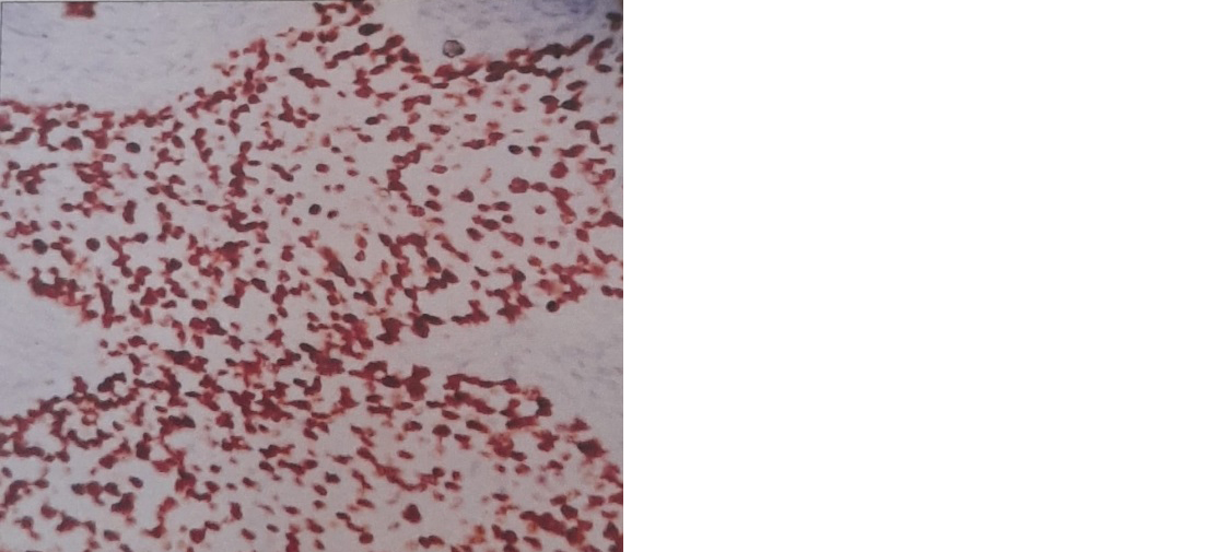
p53 expression was observed in 68% of ESCC patients. p53 expression was found higher in patients with age group ≥60 years than <60 years. There was no statistically significant correlation between gender and p53 expression profile. Higher p53 expression was found in patients who are betel nut chewers (P =0.0434). Only 38% of well differentiated tumors (Figure 2) showed positive p53 expression when compared to moderately differentiated (Figure 3) and poorly differentiated tumors (Figure 4), which showed 71% and 85% positivity respectively. So there was significant correlation between P53 expression and histological grade of cancer (P =0.0262).
There were no significant associations between p53 expression and site, lymph node metastasis (N classification) and stage.

Figure 2: Tumor cells exhibiting p53 expression in well differentiated squamous cell carcinoma (IHC; 400X).

Figure 3: Tumor cells exhibiting p53 expression in moderately differentiated squamous cell carcinoma (IHC; 400X).

Figure 4: Tumor cells exhibiting p53 expression in poorly differentiated squamous cell carcinoma.
Table 1: Correlation between P53 expression and clinicopathologic parameters.
|
Clinicopathological parameters
|
Total
|
P53 Positive N (%)
|
P53 Negative N (%)
|
P value
|
|
Male
|
35
|
25(71%)
|
10(29%)
|
0.384
|
|
Female
|
20
|
12(60%)
|
8(40%)
|
|
<60
|
29
|
14(48%)
|
12(52%)
|
0.0444
|
|
≥60
|
26
|
23(88%)
|
6(22%)
|
|
Betel nut chewer
|
29
|
16(55%)
|
13(45%)
|
0.0434
|
|
Non betel nut chewer
|
26
|
21(81%)
|
5(19%)
|
|
Upper esophagus
|
11
|
8(73%)
|
3(27%)
|
0.4331
|
|
Middle esophagus
|
34
|
24(70%)
|
10(30%)
|
|
Lower esophagus
|
10
|
5(50%)
|
5(50%)
|
|
<5cm
|
44
|
31(70%)
|
13(30%)
|
0.3145
|
|
≥5cm
|
11
|
6(54%)
|
5(45%)
|
|
Well differentiated
|
13
|
5(38%)
|
8(62%)
|
0.0262
|
|
Moderately differentiated
|
28
|
20(71%)
|
8(29%)
|
|
Poorly differentiated
|
14
|
12(85%)
|
2(15%)
|
|
Stage1-Stage 2
|
18
|
11(61%)
|
7(39%)
|
0.3175
|
|
Stage 3-Stage 4
|
6
|
5(83%)
|
1(17%)
|
|
Nodal involvement
|
7
|
6(86%)
|
1(14%)
|
0.2040
|
|
No nodal involvement
|
17
|
10(58%)
|
7(42%)
|
Discussion
Environmental as well as genetic factors play a role in tumorigenesis of ESCC [8]. The pathogenesis of ESCC remains still unclear. In many studies it was found to be multifactorial [9]. Mutations in p53 protein can promote proliferation, invasion, metastasis, and survival of tumors [10]. So the accumulation of p53 protein indicate P53 gene mutation and the protein accumulation can be detected by immunohistochemistry. In this study p53 expression was observed in 68% of ESCC patients. In the study done by Niyaz et al [7] 54.5% showed positivity. In the study done by Sankalecha et al [9] high expression of p53 was found in 61.64%.
In the present study, there was no significant correlation between p53 positivity with patient’s gender. This is consistent with study done by Yao et a1 [11] and Sankalecha et al [9]. So the p53 mutations are largely independent of sex.
Increase in age increase the risk of carcinogenesis. A study done by Cummings et al [12] and Sankalecha et al [9] confirmed that age group of >60 years has significantly higher risk for ESCC. Similar results were observed in our study that expression of p53 was significantly higher in patients with age group of >60 years. This could be due to cumulative genetic damage that occurs with aging, which increases the likelyhood of mutations in tumor suppressor gene like p53.
Betel nut chewing has been strongly associated with the development of SCC. In this study, p53 expression was observed to be significantly high in patients with the habit of betel nut chewing. Similar results were reported by Sankalecha et al [9] and Goan et al [13]. Betel nut chewing is a known risk factor for various oral and esophageal cancers due to its carcinogenic components like arecoline which induces DNA damage. This finding emphasizes the importance of public health interventions to reduce betel nut consumption. It also highlights the need for further research into the mechanisms by which betel nut constituents induce p53 mutations.
In the present study, there was no significant correlation between p53 positivity and site of tumor. Similar findings were observed by Yao et al [14]. Whether the tumor is located in upper, middle or lower esophagus, p53 expression is uniform across different tumor sites.
Histopathologic grade of tumor is one of the important prognostic factors in esophageal squamous cell carcinoma of esophagus. When grade increases, the prognosis becomes poorer. In the present study there was a significant correlation between P53 expression and histological grade of tumor. Similar results were seen by Huang et al [15]. in 2014, Yao et al [14] and Niyaz et al [7]. This suggests that as tumors become more undifferentiated and aggressive, p53 mutations play a critical role in tumor progression. So p53 expression can serve as a prognostic marker to identify patients with more aggressive forms of disease.
In this study there was no significant associations between p53 expression and TNM stage, lymph node metastasis. Similar results were seen in study done by Niyaz et al [7], Geramizadeh et al [16] (Table 2).
Table 2: Comparison of p53 expression with clinicopathological parameters in various studies.
|
Author
|
Publication year
|
Association with age (p-value))
|
Association with gender (p-value
|
Association with grade (p-value)
|
Association with tumour extension (p-value)
|
|
Niyaz et al [7]
|
2020
|
0.447
|
0.509
|
0.567
|
0.450
|
|
Geramizah et al [16]
|
2021
|
0.281
|
1
|
1
|
0.031
|
|
Sankalecha et al [9]
|
2017
|
<0.023
|
<0.481
|
<0.001
|
<0.004
|
|
Yao et al [14]
|
2014
|
0.97
|
0.06
|
< 0.001
|
-
|
Limitation: Tertiary centre data do not precisely reflect the disease profile of the community.
Conclusion
The expression of p53 in ESCC patients significantly correlates with age, betel nut chewing and histological grade. P53 gene abnormalities are not only an early event in esophageal squamous cell carcinoma, they also play an important role in tumor progression affecting the patient survival. Therefore, the search for prognostic biomarkers like P53 is warranted even in less advanced stages. Understanding the molecular alterations in p53 and their clinical significance in ESCC may lead to improved diagnostic, prognostic and therapeutic strategies, ultimately enhancing patient outcomes.
Conflicts of interest
Authors declare no conflicts of interest.
References
[1] Jemal A. Global cancer statistics 2018: GLOBOCAN estimates of incidence and mortality worldwide for 36 cancers in 185 countries. CA Cancer J Clin. 2018; 68:394–424.
[2] Sankalecha TH, Gupta SJ, Gaikwad NR, Shirole NU, Kothari HG. Yield of p53 expression in esophageal squamous cell cancer and its relationship with survival. Saudi J Gastroenterol. 2017; 23:281–286.
[3] Lin Y, Totsuka Y, He Y, Kikuchi S, Qiao Y, et al. Epidemiology of esophageal cancer in Japan and China. J Epidermoil. 2013; 23:233–242.
[4] Zhu HC, Yang X, Xu LP, Zhao LJ, Tao GZ, et al. Meat consumption is associated with esophageal cancer risk in a meat- and cancer-histological-type dependent manner. Dig Dis Sci. 2014; 59:664–673.
[5] Pennathur A, Gibson MK, Jobe BA, Luketich JD. Oesophageal carcinoma. Lancet. 2013; 381:400–412.
[6] Das M, Sharma SK, Sekhon GS, Mahanta J, Phukan RK, et al. p16 gene silencing along with p53 single-nucleotide polymorphism and risk of esophageal cancer in Northeast India. Tumor Biol. 2017; 39:1010428317698384.
[7] Niyaz M, Ainiwaer U, Abudureheman A, Zhang L, Sheyhidin I, et al. Association between TP53 gene deletion and protein expression in esophageal squamous cell carcinoma and its prognostic significance. Oncology letters. 2020; 20:1855–1865.
[8] Chattopadhyay I. A brief overview of genetics of esophageal squamous cell carcinoma. J Cell Sci Molecul Biol. 2014; 1:103.
[9] Sankalecha TH, Gupta SJ, Gaikwad NR, Shirole NU, Kothari HG. Yield of p53 expression in esophageal squamous cell cancer and its rela tionship with survival. Saudi J Gastroenterol. 2017; 23:281–286.
[10] Muller PA, Vousden KH. p53 mutations in cancer. Nat Cell Biol. 2013; 15:2–8.
[11] Yao W, Qin X, Qi B, Lu J, Guo L, et al. Association of p53 expression with prognosis in patients with esophageal squamous cell carcinoma. Int J Clin Exp Pathol. 2014; 7:7158–7163.
[12] Cummings LC, Cooper GS. Descriptive epidemiology of esophageal carcinoma in the Ohio Cancer Registry. Cancer Detect Prev. 2008; 32:87–92.
[13] Goan YG, Chang HC, Hsu HK, Chou YP, Cheng JT. Risk of p53 gene mutation in esophageal squamous cell carcinoma and habit of betel quid chewing in Taiwanese. Cancer Sci. 2005; 96:758–765.
[14] Yao W, Qin X, Bo Qi, Lu J, Guo L, et al. Association of p53 expression with prognosis in patients with esophageal squamous cell carcinoma Int J Clin Exp Pathol. 2014; 7:7158–7163.
[15] Huang K, Chen L, Zhang J, Wu Z, Lu B, et al. Elevated p53 expression levels correlate with tumor progression and poor prognosis in patients exhibiting esophageal squamous cell carcinoma. Oncology Lett. 2014; 8:1441–1446.
[16] Geramizadeh B, Mohammadian A, Shojazadeh A, Mohammadzadeh S. P16 and P53 expression in esophageal squamous cell carcinoma: a brief report from the experience of South of Iran, and review of the literature. Clin Pathol. 2021; 14:2632010X211013821.