Full Text
Introduction
Erythema nodosum (EN) is an inflammatory disorder characterized by the appearance of tender, erythematous subcutaneous nodules, most commonly located on the anterior aspects of the lower extremities. EN is the most common form of panniculitis and is characterized histologically by inflammation of the subcutaneous fat, predominantly involving the septa without vasculitis. The condition is associated with a wide range of etiologies, including infections, autoimmune disorders, medications, sarcoidosis, inflammatory bowel disease, and malignancies. Among infectious causes, tuberculosis (TB) remains an important etiological factor, particularly in regions where TB is endemic [1,2].
Tuberculosis-associated erythema nodosum is considered a manifestation of a delayed-type (Type IV) hypersensitivity reaction to mycobacterial antigens rather than a direct infection of the skin. Histopathological examination may reveal septal panniculitis with or without granulomatous inflammation, reflecting the underlying immune response to Mycobacterium tuberculosis [3].
The diagnosis of TB-associated EN can be challenging because of its nonspecific clinical presentation and the difficulty in establishing active tuberculosis in some patients. The skin lesions may be misdiagnosed as cellulitis, thrombophlebitis, or other inflammatory and vascular disorders, resulting in delayed diagnosis and treatment [4]. Therefore, a comprehensive diagnostic approach is required, including detailed clinical evaluation, laboratory investigations, tuberculin skin testing, radiological imaging, and histopathological examination. Histopathology may demonstrate granulomatous inflammation, while imaging studies may provide evidence of pulmonary or extrapulmonary tuberculosis [5,6].
Early recognition of tuberculosis as the underlying cause of erythema nodosum is essential because appropriate antitubercular therapy can lead to complete resolution of the lesions and prevent disease progression. Herein, we report the case of a 49-year-old woman with recurrent painful erythematous nodules over both lower limbs who was initially misdiagnosed as having thrombophlebitis and cellulitis but was subsequently found to have tuberculosis-associated erythema nodosum. This case highlights the importance of considering tuberculosis in the differential diagnosis of chronic or recurrent panniculitis, particularly in TB-endemic regions [7].
Case presentation
A 49-year-old woman presented to the Rheumatology Department with complaints of recurrent painful skin nodules associated with a burning sensation over the medial aspects of both legs for the past six years. She denied any constitutional or systemic symptoms, including fever, cough, dysuria, fatigue, malaise, arthralgia, oral ulcers, sore throat, sinusitis, or weight loss. However, she reported excessive hair loss. She had previously consulted multiple physicians and had initially been diagnosed with thrombophlebitis and cellulitis. Treatment with compression stockings (Grade 2), Daflon® 500 mg twice daily for thrombophlebitis, and Chymoral Forte for cellulitis resulted in symptomatic improvement for approximately six months; however, the lesions subsequently recurred. Owing to the persistent and recurrent nature of her symptoms, she was referred to our department for further evaluation in collaboration with dermatology.
On physical examination, the patient appeared pale. Multiple erythematous, tender subcutaneous nodules were observed over the anterior aspects of both tibiae, a characteristic distribution of erythema nodosum (Figure 1). A larger nodule was present on the right leg, whereas two smaller nodules were noted on the left leg. Dilated superficial veins were observed bilaterally. Her vital signs were stable, with a blood pressure of 130/70 mmHg, respiratory rate of 20 breaths/min, and oxygen saturation of 98% on room air. The remainder of the systemic examination, including cardiovascular, respiratory, musculoskeletal, and neurological assessments, was unremarkable.
Routine laboratory investigations were largely within normal limits except for elevated high-sensitivity C-reactive protein (hs-CRP) levels of 1.4 mg/L (reference range: <0.1 mg/L) and angiotensin-converting enzyme (ACE) levels of 81.64 nmol/mL/min (reference range: <52 nmol/mL/min). The Mantoux test was strongly positive, with an induration measuring 15 mm. Autoimmune screening, including rheumatoid factor and anti-double-stranded DNA (anti-dsDNA) antibodies, was negative. Human leukocyte antigen (HLA)-B51 testing was also negative.
Contrast-enhanced computed tomography (CECT) of the chest and abdomen demonstrated two enlarged non-necrotic right lower cervical lymph nodes and multiple tiny subpleural nodules involving the right middle lobe and the lateral basal segments of both lower lobes. In addition, a hypodense nodule with homogeneous hypoenhancement was identified in the left lobe of the thyroid gland. Excisional biopsy of a cervical lymph node was performed, and histopathological examination revealed necrotizing granulomatous inflammation, findings highly suggestive of tuberculosis (Figure 2). Ziehl–Neelsen staining for acid-fast bacilli (AFB) was negative, and AFB cultures remained negative.
Based on the clinical presentation of recurrent erythematous nodules, a strongly positive Mantoux test, radiological evidence of pulmonary involvement, and histopathological findings of necrotizing granulomatous inflammation, a diagnosis of tuberculosis-associated erythema nodosum was established. The patient was subsequently evaluated by an infectious disease specialist, who confirmed the diagnosis of tuberculosis. She was initiated on standard first-line antitubercular therapy consisting of isoniazid, rifampicin, pyrazinamide, and ethambutol.
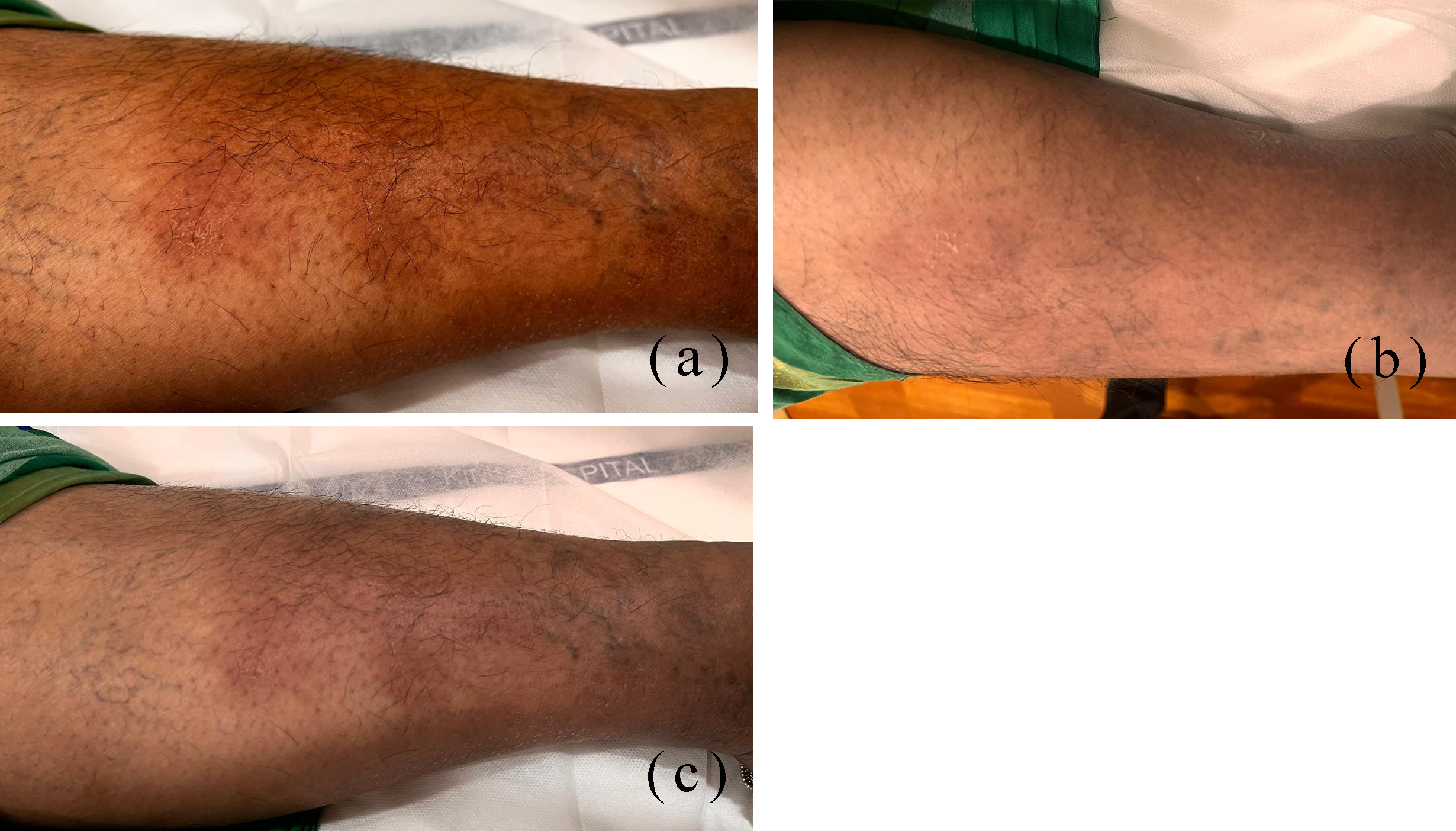
Figure 1: Clinical photographs (a–c) demonstrating multiple erythematous, tender subcutaneous nodules over the anterior aspects of both tibiae, consistent with erythema nodosum.
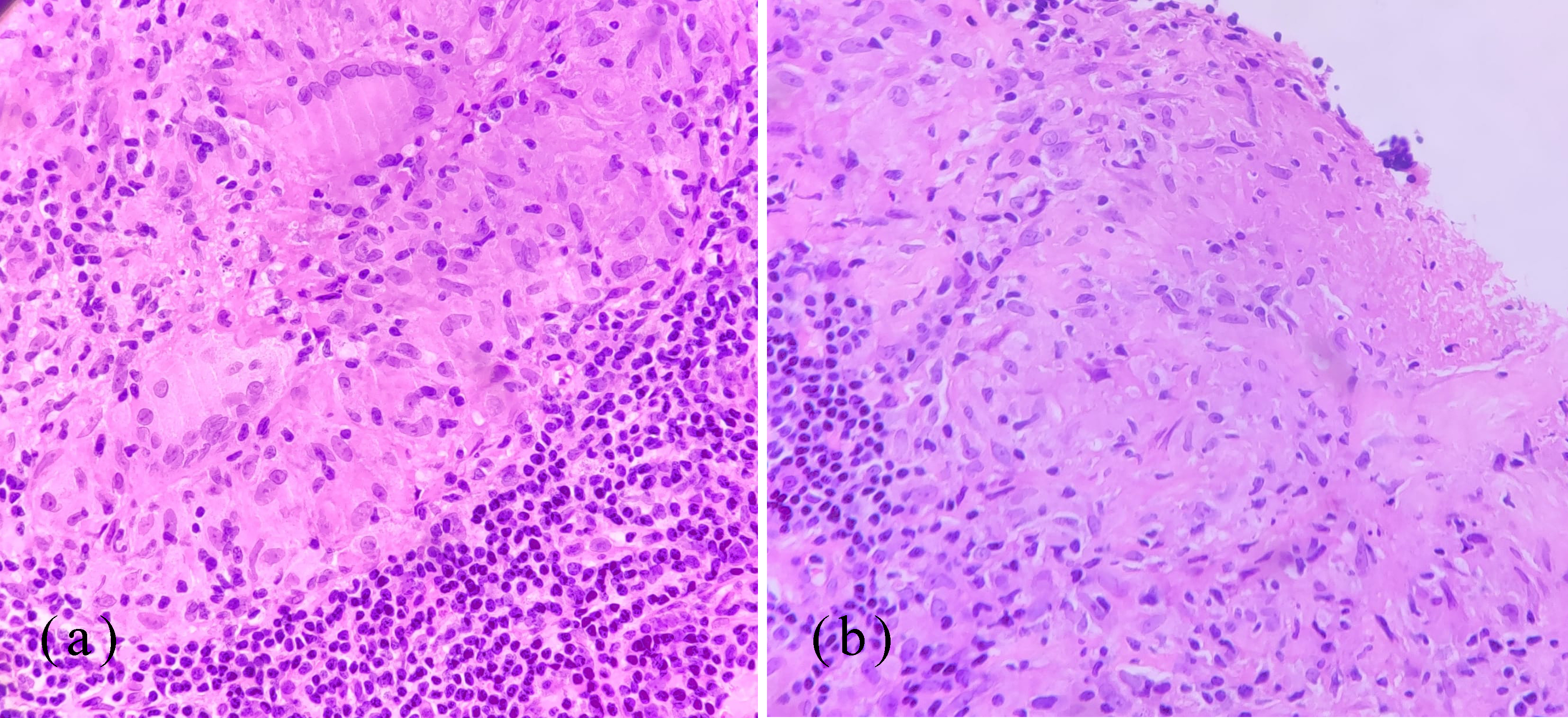
Figure 2: Histopathological findings of cervical lymph node biopsy showing features consistent with tuberculosis. (a) High-power photomicrograph demonstrating dense infiltration of lymphocytes and epithelioid histiocytes with occasional Langhans-type giant cells, consistent with granulomatous inflammation associated with tuberculosis. (b) Low-power photomicrograph showing well-formed granulomas with central caseous necrosis surrounded by a peripheral rim of lymphocytic infiltrate, characteristic of tuberculosis.
Discussion
Erythema nodosum (EN) is the most common form of panniculitis and is frequently associated with a variety of underlying conditions, including infections, autoimmune disorders, medications, and malignancies. Among infectious causes, tuberculosis (TB) remains an important etiological factor, particularly in regions where TB is endemic. EN associated with TB is generally considered a delayed-type (Type IV) hypersensitivity reaction to Mycobacterium tuberculosis antigens and typically manifests as painful, erythematous nodules over the lower extremities [7]. The present case of a 49-year-old woman with recurrent EN in the absence of significant constitutional or respiratory symptoms illustrates the diagnostic challenge posed by TB-associated EN, particularly in patients with subclinical or extrapulmonary disease [8].
The patient was initially diagnosed with thrombophlebitis and cellulitis and received symptomatic treatment with temporary improvement. However, the recurrence of painful nodules over the shins, a characteristic site for EN, prompted further evaluation. Similar cases have been reported in which the diagnosis of tuberculosis was delayed because of the absence of classical pulmonary manifestations, emphasizing the importance of considering TB in the differential diagnosis of recurrent or persistent EN, especially in endemic areas [8].
In the present case, the patient demonstrated several findings suggestive of tuberculosis, including a strongly positive Mantoux test, elevated inflammatory markers, radiological evidence of cervical lymphadenopathy and pulmonary nodules, and histopathological findings of necrotizing granulomatous inflammation with Langhans-type giant cells. Although Ziehl–Neelsen staining and AFB cultures were negative, these results do not exclude tuberculosis, as microbiological confirmation may be lacking in a substantial proportion of extrapulmonary TB cases [9].
An important differential diagnosis in this case was sarcoidosis, particularly because the patient had elevated serum angiotensin-converting enzyme (ACE) levels and granulomatous inflammation on histopathology. Necrotizing sarcoid granulomatosis (NSG), a rare variant of sarcoidosis, may occasionally exhibit necrosis and mimic tuberculosis both clinically and histologically [10]. However, sarcoidosis is classically characterized by non-caseating granulomas, and when necrosis is present, it is usually focal and limited. In contrast, the extensive necrotizing granulomatous inflammation observed in the present case, together with a strongly positive Mantoux test and radiological findings suggestive of tuberculosis, favored TB as the underlying diagnosis [11].
The coexistence of elevated ACE levels and necrotizing granulomas posed a diagnostic dilemma because these findings may be encountered in both tuberculosis and sarcoidosis. Furthermore, Langhans-type giant cells are not specific to TB and may also be observed in sarcoidosis and other granulomatous disorders. Therefore, the diagnosis of granulomatous diseases should rely on a comprehensive assessment integrating clinical presentation, laboratory investigations, imaging findings, histopathology, and epidemiological context rather than on a single diagnostic feature [12].
Considering the high prevalence of tuberculosis in endemic regions and the overall clinical, radiological, and histopathological findings, antitubercular therapy was initiated, resulting in significant clinical improvement. The favorable response to treatment further supported the diagnosis of tuberculosis-associated erythema nodosum [13].
This case highlights the complex diagnostic overlap between tuberculosis and sarcoidosis and underscores the importance of maintaining a high index of suspicion for TB in patients presenting with recurrent erythema nodosum, even in the absence of classical pulmonary symptoms. Early diagnosis and prompt initiation of appropriate therapy are essential to prevent disease progression and achieve complete resolution of the associated cutaneous manifestations [14].
Conclusion
This case highlights tuberculosis as an important underlying cause of erythema nodosum, particularly in endemic regions and in patients presenting with recurrent skin nodules without overt systemic symptoms. The diagnostic overlap with other granulomatous and inflammatory conditions necessitates a comprehensive evaluation incorporating clinical, laboratory, radiological, and histopathological findings. Early recognition of tuberculosis-associated erythema nodosum and prompt initiation of antitubercular therapy are crucial for achieving clinical resolution, preventing disease progression, and improving patient outcomes.
Conflicts of interest
Authors declare no conflicts of interest.
References
[1] Rizvi Z, Iqbal T, Javed A, Rizvi A. Erythema nodosum: a consequence of tuberculosis. Cureus. 2019; 11.
[2] Perez–Garza DM, Chavez–Alvarez S, Ocampo–Candiani J, Gomez–Flores M. Erythema nodosum: a practical approach and diagnostic algorithm. Am J Clin Dermatol. 2021; 22:367–378.
[3] Kritsotakis E. Erythema nodosum as sign of primary tuberculosis. Oxford Med Case Reports. 2017; 8:041.
[4] Meienberger N, Maul JT, Frohlich F, Maul LV, Kundig T, et al. Atypical and typical presentation of erythema nodosum: clinical differences in treatment and outcome. Dermatology. 2024; 240:226–232.
[5] Laborada J, Cohen PR. Tuberculosis–associated erythema nodosum. Cureus. 2021; 13.
[6] Chen S, Chen J, Chen L, Zhang Q, Luo X, et al. Mycobacterium tuberculosis infection is associated with the development of erythema nodosum and nodular vasculitis. PLoS One. 2013; 8.
[7] Rodriguez–Takeuchi SY, Renjifo ME, Medina FJ. Extrapulmonary tuberculosis: pathophysiology and imaging findings. Radiographics. 2019; 39:2023–2037.
[8] Lee JY. Diagnosis and treatment of extrapulmonary tuberculosis. Tuberc Respir Dis (Seoul). 2015; 78:47–55.
[9] Mert A, Ozaras R, Tabak F, Ozturk R. Primary tuberculosis cases presenting with erythema nodosum. J Dermatol. 2004; 31:66–68.
[10] Ali AS, Abdullah BH. Granulomatous Lesions in the Head and Neck Region: A Clinicopathological, Histochemical, and Molecular Diagnostic Study. Diagnostics (Basel). 2025;15(16):2055.
[11] Yang G, Tan Z, Zhou L, Yang M, Peng L, et al. Effects of angiotensin II receptor blockers and ACE (angiotensin–converting enzyme) inhibitors on virus infection, inflammatory status, and clinical outcomes in patients with COVID–19 and hypertension. Hypertension. 2020; 76:51–58.
[12] Sarcoid-like Reaction in Breast Cancer Tumor Bed and Axillary Lymph Nodes Following Neoadjuvant Chemotherapy: A Case Report. Ann Clin Lab Sci. 2025 Mar;55(2):281-285.
[13] Mukhopadhyay S, Gal AA. Granulomatous lung disease: an approach to the differential diagnosis. Arch Pathol Lab Med. 2010; 134:667–690.
[14] Judson MA. The diagnosis of sarcoidosis. Clin Chest Med. 2008; 29:415–427.