Orginal Research
2023
June
Volume : 11
Issue : 2
Tibiotalocalcaneal arthrodesis with retrograde intramedullary nail for Charcot neuroarthropathy of foot - A prospective observational study
Shan AK, Binoy S, Biju S, Kumar KA, Shibu R
Pdf Page Numbers :- 131-135
Shan AK1, Binoy S1, Biju S1, Akhil Kumar K1, and Shibu R1,*
1Department of Orthopaedics, Government Medical College, Thiruvananthapuram, Kerala-695011, India
*Corresponding author: Dr. R. Shibu, Assistant Professor, Department of Orthopaedics, Government Medical College, Thiruvananthapuram, Kerala-695011, India. Email: drshibur@gmail.com
Received 9 January 2023; Revised 8 March 2023; Accepted 15 March 2023; Published 23 March 2023
Citation: Shan AK, Binoy S, Biju S, Kumar KA, Shibu R. Tibiotalocalcaneal arthrodesis with retrograde intramedullary nail for Charcot neuroarthropathy of foot - A prospective observational study. J Med Sci Res. 2023; 11(2):131-135. DOI: http://dx.doi.org/10.17727/JMSR.2023/11-25
Copyright: © 2023 Shan AK et al. Published by KIMS Foundation and Research Center. This is an open-access article distributed under the terms of the Creative Commons Attribution License, which permits unrestricted use, distribution, and reproduction in any medium, provided the original author and source are credited.
Abstract
Background: Surgical treatment is often needed for Charcot neuroarthropathy (CN) of foot. Even though good outcomes were reported with tibiotalocalcaneal arthrodesis (TTCA) in CN patients, the choice of implant is still an issue. The aim of the study was to evaluate the outcomes of TTCA with retrograde intramedullary nail, in patients with CN.
Materials and methods: A prospective observational study was done with 41 consecutive patients treated with TTCA with hindfoot retrograde IM nail. The American Orthopaedic Foot and Ankle Society (AOFAS) score was used for the outcome evaluation, one year after surgery.
Results: The mean age was 67.4(±8.7) years, with 63.5% females. Most common cause was diabetes mellitus (75.6%), followed by post-traumatic CN (19.5%) and chronic alcoholism (4.9%). Eleven (26.8%) had good score at the end of 1 year compared to five (12.2%) pre-op and twenty (48.8%) had fair score compared to fifteen (36.6%). The AOFAS score increased one year after surgery (p value 0.001). In twenty (48.8%) patients, the time taken for union was more than 6 months; and in 15 (36.6%) union occurred within 6 months of surgery. Six (14.6%) patients had non-union. Two (4.9%) had deep infection along with non-union. Hardware failure was seen in 3 (7.3%) patients. Hardware failure with non-union was reported in one (2.4%). Four (9.8%) patients had superficial wound infection and amputation done in one (2.4%) patient.
Conclusion: Tibiotalocalcaneal arthrodesis with retrograde intramedullary nail is an acceptable and safe procedure with good clinical outcomes in patients with Charcot arthropathy which obviates the need for amputation.
Keywords: Charcot neuroarthropathy; tibiotalocalcaneal arthrodesis; intramedullary nail; amputation
Full Text
Introduction
Charcot neuroarthropathy (CN) of foot was first described by Jean Martin Charcot in 1868 in association with tabes dorsalis resulting from tertiary syphilis. Jordan in 1936, first noted its relationship to diabetes, which is now recognized as the main cause of Charcot foot. It develops in 1 per 600 diabetic patients and in 1 per 100 patients with diabetic neuropathy [1-3]. Since the pathophysiology of CN is unclear, several theories were proposed [4]. According to neurotraumatic hypothesis, CN results from loss of protective sensation leading to microtrauma of foot. As per the neurotrophic hypothesis, sympathetic neuropathy leads to increased blood flow to the limb resulting in soft tissue swelling, increased susceptibility to osteoclast-mediated bone resorption and progressive fractures and dislocations. As the disease progress, the plantar arch collapses and the increased pressure on the plantar osseous prominences along with the decreased plantar sensation results in plantar ulcerations. This leads to deep soft tissue infection and osteomyelitis ultimately resulting in amputation. Around 40- 60% of all amputations of the lower limbs are done on patients with diabetic Charcot foot [5-7]. Less frequently CN is associated with syringomyelia, alcoholism and leprosy.
Charcot foot presents an important diagnostic and therapeutic challenge to clinicians. Some patients can be treated with conservative management in the active acute stage. Offloading the foot and immobilization with an irremovable total contact cast (TCC) is done to prevent further destruction and to preserve the structure of foot. Longer duration of TCC leads to osteoarthritis [8, 9]. Surgical treatment is often needed for the management of deformities and ulcerations of chronic Charcot foot. Exostectomy, external or internal arthrodesis and amputation are done depending on the type of deformity and clinical condition of the patients [10].
Even though good clinical outcomes were reported by many authors regarding arthrodesis in CN patients, the choice of most appropriate implant for arthrodesis is still an issue [11]. Tibiotalocalcaneal arthrodesis (TTCA) can be done to save CN patients from amputation [12]. It can be done with plate and screws, screws alone, external fixators or with retrograde nail. Dalla Paola L et al. reported good outcome from TTCA done with retrograde interlocking nails in Charcot arthropathy without ulcerations [13]. There are several studies regarding TTCA using various implants, but only few studies with TTCA done with hindfoot retrograde intramedullary (IM) locking nails. The aim of the present study was to report the outcome of TTCA done with hind foot retrograde intramedullary nail, in patients with Charcot joint, after a minimum follow up of one year using the American Orthopaedic Foot and Ankle Society (AOFAS) Ankle-Hindfoot score [14].
Materials and methods
The study was conducted as a prospective observational study at Government Medical College, Thiruvananthapuram during the period June 2021 to November 2022 after obtaining the institutional Ethics Committee clearance. All consecutive patients with Charcot arthropathy who were managed by tibiotalocalcaneal arthrodesis with hindfoot retrograde nail in the institution were included in the study.
The inclusion criteria were- patients in the age group 40-90 years of either sex, who had Charcot arthropathy of ankle and subtalar joint, and who gave informed consent for the study. The exclusion criteria were- patients with previous disability, patients with severe comorbidities who were unfit for surgery, uncontrolled diabetes mellitus, children, associated neurovascular injuries and patients who were lost to follow-up.
There was a total of 41 patients in the study. Complete clinical and neurological examination were done for all patients. Radiographs of ankle in antero-posterior (AP), lateral and mortise views; and foot – AP and lateral views were taken for all patients. The AOFAS Ankle-Hindfoot score was used for the outcome evaluation of ankle and hind foot. The AOFAS score is the most used score for measuring the outcome of treatment in patients with complex ankle or hindfoot injury. It combinedly evaluates clinician reported and patient reported parts [14]. Both pre-op and post-op AOFAS scores were calculated. Assessment was done at 1 month, 3 months, 6 months and 1 year after surgery. The AOFAS score at 1 year was considered for evaluation. An AOFAS score >75 out of 100 implies good functional outcome, AOFAS score of 50-75 implies fair outcome and AOFAS score of <50 implies poor outcome. Complications including infection, non-union and hardware failure etc were also noted.
Statistical analysis
The collected data was entered into Excel sheet. Categorical variables were expressed as proportions and quantitative variables as mean and standard deviation. Outcome of the subjects were analysed with clinical and functional aspects and indices calculated. Data analyses were done using SPSS Statistics version 22.0 (IBM Corp; Chicago, USA). P value of <0.05 was considered statistically significant.
Surgical procedure
All cases were done under spinal anaesthesia in supine position. Correction of deformity was done through an extended lateral approach to the ankle and subtalar joint. The ankle joint and subtalar joints were prepared for arthrodesis along with correction of deformity. The entry point for the nail at heel was the point of intersection of a line from the second toe to the centre of the heel and another line at the junction of the anterior and middle thirds of the heel pad. A guide wire was passed through the centre of medullary canal of tibia, under image intensification. Reaming was done with sequential reamers over the guide wire, through a tissue protector. Then an appropriately sized intramedullary locking nail was introduced into the medullary canal. Before final seating of the nail, bone graft from the morselized malleoli was placed in the arthrodesis site and in the sinus tarsi area of the calcaneus. The position of the nail and position of TTCA were re-evaluated using image intensification and locking screws applied. The position of ankle arthrodesis was neutral in sagittal plane, neutral to 5º valgus in coronal plane, 5-10o external rotation in horizontal plane, and talus exactly below the tibia. Post-operatively a short leg splint was applied with ankle in neutral position. After 2 weeks, the splint was replaced with a short leg non-weight bearing cast for another 4 weeks. Patients were then advanced to full weight bearing in a walking boot over 4-6 weeks. Patients were followed up for a minimum of one year (Figures 1 and 2).

Figure 1: Pre-op X-ray- Charcot joint with destruction of ankle.
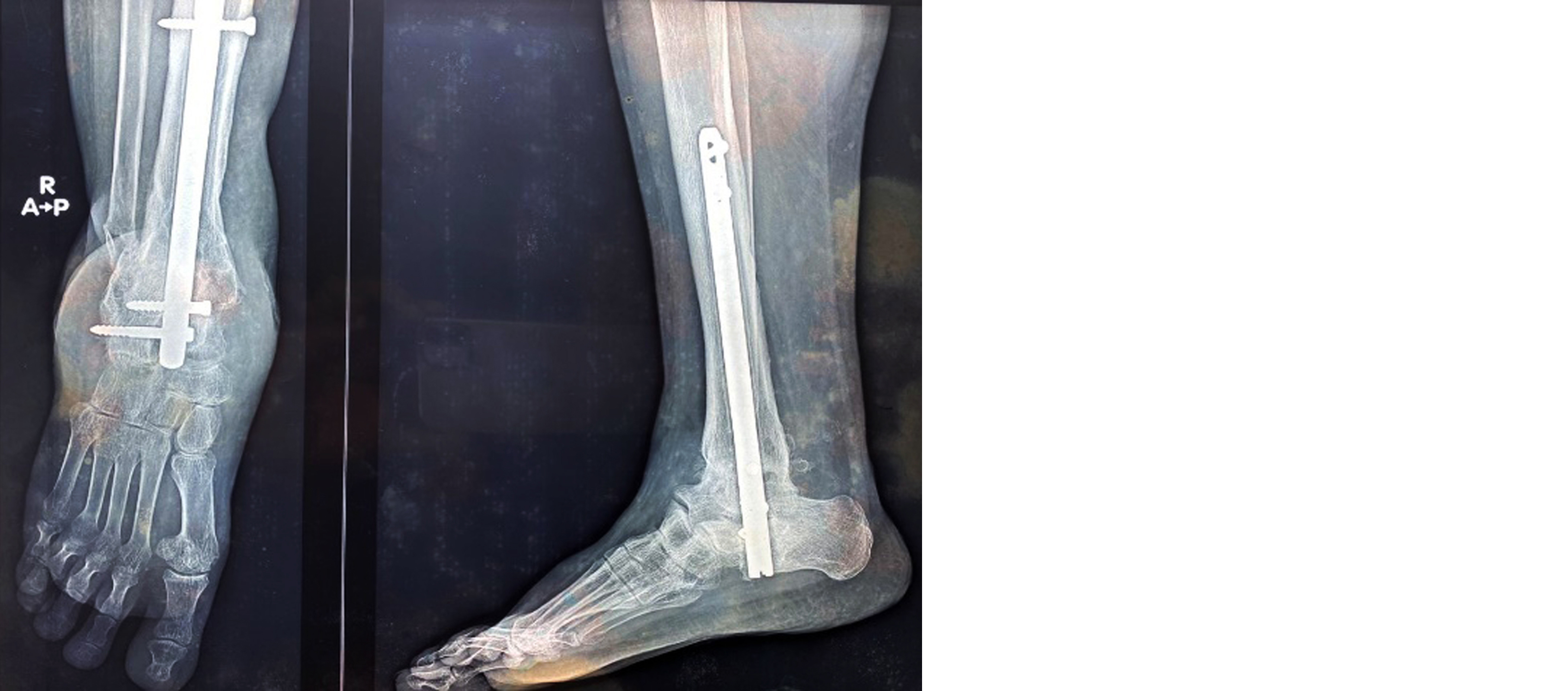
Figure 2: Post-op X-ray. United ankle arthrodesis with IM nail in situ.
Results
The mean age of the 41 patients was 67.4 (±12.7) years, with 25 (61%) belonging to 61-75 years. Twenty-six (63.5%) patients were females. Most common cause was diabetes mellitus (75.6%), followed by post-traumatic CN 19.5% and chronic alcoholism 4.9%. Fifteen (36.5%) had other comorbidities as well (Table 1).
Table 1: Age and gender distribution of the study participants (N=41).
|
Characteristics
|
Frequency (%)
|
|
Age group
|
|
| |
<60 years
|
11 (26.8)
|
| |
61-75 years
|
25 (61.0)
|
| |
>75 years
|
5 (12.2)
|
|
Sex
|
|
|
| |
Male
|
15 (36.5)
|
| |
Female
|
26 (63.5)
|
| |
Etiology
|
|
| |
Chronic Alcoholism
|
2 (4.9)
|
| |
Diabetes Mellitus
|
31 (75.6)
|
| |
Post Traumatic
|
8 (19.5)
|
|
Other comorbidities
|
| |
Yes
|
15 (36.5)
|
| |
No
|
26 (63.5)
|
Eleven (26.8%) of the study participants had good score at the end of 1 year compared to five (12.2%) pre-op. Twenty (48.8%) had fair score at 1 year compared to fifteen (36.6%). Ten patients (24.4%) had poor score at 1 year, compared to 21 (51.2%) prior to surgery. The increase in AOFAS score one year after surgery was found to be statistically significant (p value=0.001) (Table 2).
Table 2: Distribution of AOFAS score among the study participants (N=41).
|
AOFAS score
|
Preop AOFAS score
|
Postop AOFAS score (1 year)
|
P value*
|
|
Good (>75)
|
5 (12.2)
|
11 (26.8)
|
<0.001
|
|
Fair (50-75)
|
15 (36.6)
|
20 (48.8)
|
|
Poor (<50)
|
21 (51.2)
|
10 (24.4)
|
*P value <0.05- statistically significant.
In twenty (48.8%) patients, the time taken for union was more than 6 months; and in 15 (36.6%) union occurred within 6 months of surgery. Six (14.6%) patients had non-union. Thirteen (31.7%) patients reported some or other complications. Two (4.9%) had deep infection along with non-union. Hardware failure was seen in 3 (7.3%) patients. Hardware failure with non-union was reported in one (2.4%). Non-union without infection and hardware failure was seen in 3 patients (7.3%). Four (9.8%) patients had superficial wound infection. Infections were controlled with parenteral antibiotics in 5 of the six patients with infection. In one patient (2.4%), infection was not settled with antibiotics and had to undergo below knee amputation (Table 3).
Table 3: Distribution complications and its outcome among the participants (N=41).
|
Characteristics
|
Frequency (%)
|
|
Complications
|
|
|
Yes
|
13 (31.7)
|
|
|
No
|
28 (68.3)
|
|
Type of complication
|
|
|
Deep infection & Non-union
|
2 (4.9)
|
|
|
Hardware failure
|
3 (7.3)
|
|
|
Non-union
|
3 (7.3)
|
|
|
Superficial wound infection
|
4 (9.8)
|
|
|
Hardware failure, non-union
|
1 (2.4)
|
|
Treatment for infections
|
|
|
Controlled with IV antibiotics
|
5 (12.2)
|
|
|
Not settled with antibiotics and underwent BK* amputation
|
1 (2.4)
|
|
Time for fusion
|
|
|
6 months and less
|
15 (36.6)
|
|
|
More than 6 months
|
20 (48.8)
|
|
|
Non-united
|
6 (14.6)
|
Note: *BK amputation - below knee amputation.
Discussion
The result of the study shows that TTCA with retrograde IM nail in Charcot ankle and hind foot deformity can make independent mobilisation of the patients possible, resulting in salvage of the limb in majority of patients. Majority of the patients in the study were in the age group of 61- 75 years with mean age 67.4 (±12.7). The AOFAS score increased one year after surgery compared to pre-op score (p value 0.001), which are comparable with other studies [12, 13, 15]. 26.8% of the patients had good and 48.8% had fair AOFAS scores one year after TTCA with IM nails, compared to a pre-operative score of 12.2% and 36.6% respectively. Twenty-eight (68.3%) patients had no complications following surgery.
The most common complication was non-union which occurred in 14.6% (n=6) patients, out of which 4.9% (n=2) had associated deep infection and 2.4% (n=1) had associated hardware failure. One patient with deep infection and non-union did not respond to antibiotic treatment and had undergone below-knee amputation. Four (9.8%) patients developed superficial infection and were controlled with two weeks of parenteral antibiotics. Chraim et al., in their study reported 26% of minor and 15% of major complications in their study of hind foot arthrodesis with retrograde IM nail in CN [16]. Patients with non-union and hardware failure were managed with custom made ankle-foot orthosis. The relatively high rate of infection may be explained by the fact that the major cause of CN in the study (75.6%) was due to diabetes mellitus. Decreased vascularity and the associated loss of protective sensation in diabetic neuropathy may be the cause of non-union and implant failure [17].
The treatment of CN includes TTCA with retrograde IM nail, external fixator, plate and/or screws or a combination of these [18, 19]. ElAlfy et al., compared external fixator with IM nailing in CN and reported the complications were higher among external fixator group [20]. Kaissar et al; reported a higher rate of fusion and lesser complications in IM nail group than external fixator group [21]. There is high incidence of pin tract infections, risk of tibia fracture and need for second surgery for implant removal in external fixation. TTCA done with locking plate fixation is associated with high failure rate, due to excessive construct rigidity [22]. Complications were higher when simultaneous midfoot fusion was done along with TTCA [15, 23].
Compared to other forms of fixation, a retrograde IM nail is believed to provide adequate stability and resist the multiplanar forces exerted by the long lever arm of foot across the ankle joint. IM nail is a load sharing device and allow early weight-bearing. It also allows axial compression at ankle and subtalar joints intra-operatively, leading to optimal bone apposition and improved bone fusion [24]. Performing TTCA with IM nail in the early chronic stage of CN, instead of postponing surgery to later stages, reduces the chance of progressive deformation which leads to ulceration and osteomyelitis [25]. Close monitoring by a multi-disciplinary team is essential in these complex patients to avoid complications. The limb salvage using IM nail may be time consuming and appear expensive compared to amputation, but Gil et al., have found no cost difference between the groups [26].
Even though our study was a prospective study with relatively large number (41) of patients, the study is limited by the lack of a control group for comparison. A prospective, multicentric randomised controlled trial may shed more light on this method.
Conclusion
Tibiotalocalcaneal arthrodesis with retrograde hind foot intramedullary nail is an acceptable and safe procedure with good clinical outcomes and acceptable complication rates in patients with Charcot arthropathy. Majority of patients achieved independent mobilisation and an improvement in the quality of life. This obviates the need for below knee amputation in majority of patients. Close monitoring by a multi-disciplinary team is essential in these complex patients to avoid complications.
Conflicts of interest
Authors declare no conflicts of interest.
References
[1] Ersin M, Demirel M, Chodza M, Bilgili F, Kilicoglu OI. Mid-term result of hindfoot arthrodesis with a retrograde intramedullary nail in 24 patients with diabetic Charcot neuroarthropathy. J Acta Orthopedica. 2020; 91:336–340.
[2] Rosskopf AB, Loupatatzis C, Pfirrmann CWA, Boni T, Berli MC. The Charcot foot: a pictorial review. Insights Imaging. 2019; 10:77.
[3] Sponer P, Kucera T, Brtková J, Srot J. The management of Charcot midfoot deformities in diabetic patients. Acta Medica (Hradec Kralove). 2013; 56:3–8.
[4] Kuharajan R, Yazid BM, Ohnmar H, Yuliawiratman BS. Functional outcome of hindfoot arthrodesis in Charcot arthropathy. Med Health. 2019; 14:172–182.
[5] Cianni L, Bocchi MB, Vitiello R, Greco T, De Marco D, et al. Arthrodesis in Charcot foot: a systematic review. Orthop Rev. 2020; 12:8670.
[6] Wukich DK, Raspovic KM, Hobizal KB, Rosario B. Radiographic analysis of diabetic midfoot Charcot neuroarthropathy with and without midfoot ulceration. Foot Ankle Int. 2014; 35:1108–1115.
[7] Strotman PK, Reif TJ, Pinzur MS. Charcot arthropathy of the foot and ankle. Foot Ankle Int. 2016; 37:1255–1263.
[8] Griffiths DA, Kaminski MR. Duration of total contact casting for resolution of acute Charcot foot: a retrospective cohort study. J Foot Ankle Res. 2021;14:44.
[9] Christensen TM, Gade-Rasmussen B, Pedersen LW, Hommel E, Holstein PE, et al. Duration of off-loading and recurrence rate in Charcot osteoarthropathy treated with less restrictive regimen with removable walker. J Diabetes Complicat. 2012; 26:430–443.
[10] Galli M, Scavone G, Vitiello R, Flex A, Caputo S et al. Surgical treatment for chronic Charcot neuroarthropathy. Foot (Edinb). 2018; 36:59–66.
[11] Burns PR, Dunse A. Tibiotalocalcaneal arthrodesis for foot and ankle deformities. Clin Podiatr Med Surg. 2017; 34:357–380.
[12] Lee BH, Fang C, Kunnasegaran R, Thevendran G. Tibiotalocalcaneal arthrodesis with the hindfoot arthrodesis nail: a prospective consecutive series from a single institution. J Foot Ankle Surg. 2018; 57:23–30.
[13] Paola LD, Volpe A, Varotto D, Brocco E, Senesi A, et al. Use of a retrograde nail for ankle arthrodesis in Charcot neuroarthropathy: a limb salvage procedure. Foot Ankle Int. 2007; 28:967–970.
[14] Lieshout EMMV, Boer ASD, Meuffels DE, Meuffels DE, Hoed PTD, et al. American Orthopaedic Foot and Ankle Society (AOFAS) AnkleHindfoot Score: a study protocol for the translation and validation of the Dutch language version. BMJ Open 2017;7:e012884.
[15] Siebachmeyer M, Boddu K, Bilal A, Hester TW, Hardwick T, et al. Outcome of one-stage correction of deformities of the ankle and hindfoot and fusion in Charcot neuroarthropathy using a retrograde intramedullary hindfoot arthrodesis nail. Bone Joint J. 2015; 97:76–82.
[16] Chraim M, Krenn S, Alrabai HM, Trnka HJ, Bock P. Mid-term follow-up of patients with hindfoot arthrodesis with retrograde compression intramedullary nail in Charcot neuroarthropathy of the hindfoot. Bone Joint J. 2018; 100:190–196.
[17] Bajuri MY, Ong SL, Das S, Mohamed IN. Charcot neuroarthropathy: current surgical management and update. A systematic review. Front Surg. 2022; 9:820826.
[18] Vopat ML, Nentwig MJ, Chong ACM, Agan JL, Shields NN, et al. Initial diagnosis and management for acute Charcot neuroarthropathy. Kans J Med. 2018; 11:114–119.
[19] Schneekloth BJ, Lowery NJ, Wukich DK. Charcot neuroarthropathy in patients with diabetes: an updated systematic review of surgical management. J Foot Ankle Surg. 2016; 55:586e590.
[20] ElAlfy B, Al AM, Fawzy SI. Ilizarov external fixator versus retrograde intramedullary nailing for ankle joint arthrodesis in diabetic Charcot neuroarthropathy. J Foot Ankle Surg. 2017; 56:309–313.
[21] Kaissar Y, Chahine A. Intramedullary nail versus external fixator for ankle arthrodesis in Charcot neuroarthropathy: A meta-analysis of comparative studies. J Ortho Surg. 2019; 27:1–7.
[22] Smith K, Araoye I, Jones C, Shah A. Outcomes of locking-plate fixation for hindfoot fusion procedures in 15 patients. J Foot Ankle Surg. 2017; 56:1188–1193.
[23] Butt DA, Hester T, Bilal A, Edmonds M, Kavarthapu V. The medial column Synthes Midfoot Fusion Bolt is associated with unacceptable rates of failure in corrective fusion for Charcot deformity: results from a consecutive case series. Bone Joint J. 2015; 97:809–813.
[24] Kavarthapu V. Surgical management: internal stabilisation. In: Edmonds M, Sumpio B, eds. Limb Salvage of the Diabetic Foot- an Interdisciplinary Approach. Springer; 2019; pp.173–184.
[25] Oesmana I, Asdib ARB. Calcaneotalotibial arthrodesis by retrograde intramedullary nailing using expert tibia nail for charcot osteoneuropathy of the foot: A case series. Int J Surg Case Rep. 2019; 57:9–14.
[26] Gil J, Schiff AP, Pinzur MS. Cost comparison: limb salvage versus amputation in diabetic patients with charcot foot. Foot Ankle Int. 2013; 34:1097–1099.