Full Text
Introduction
Cervical squamous cell carcinoma (SCC) is the fourth most common malignancy among women worldwide and remains a significant public health burden, particularly in low- and middle-income countries. In India, cervical cancer accounts for a substantial proportion of cancer-related morbidity and mortality among women, with SCC constituting approximately 70–80% of all cervical malignancies [1-3]. The tumour typically spreads through lymphatic and hematogenous routes and invades adjacent pelvic structures in advanced stages [2].
An unusual pattern of spread, termed superficial or horizontal spread, has been increasingly recognized in rare cases of cervical SCC. In this phenomenon, tumour cells extend proximally along the mucosal surface of the endometrium and may occasionally infiltrate the myometrium and adnexal structures [3, 4]. Endometrial involvement is the most frequently reported manifestation, whereas extension to the fallopian tubes, ovaries, omentum, and other extrauterine sites remains exceedingly uncommon [1,5,6].
The pathogenesis of this rare tumor spread remains poorly understood. Proposed mechanisms include clonal expansion of neoplastic cells along the epithelial lining, persistent human papillomavirus (HPV) infection, endometrial senescence, prolonged estrogen exposure, and cervical interventions such as loop electrosurgical excision procedure (LEEP) or conization [1,5]. Accurate distinction between primary adnexal neoplasms and metastatic cervical SCC is essential because the latter may mimic primary tumours both clinically and morphologically [2,7].
Herein, we report a rare case of cervical SCC exhibiting horizontal spread to the uterine corpus, bilateral fallopian tubes, and periadnexal tissue, thereby contributing to the limited literature on this unusual clinicopathological entity.
Case presentation
A cervical biopsy performed in a 60-year-old postmenopausal woman presenting with postmenopausal bleeding demonstrated HSIL. MRI of the abdomen and pelvis showed an enhancing lesion involving the anterior cervical wall with associated cervical stenosis, suggestive of an underlying invasive cervical malignancy. Based on the clinicoradiological findings, total abdominal hysterectomy with bilateral salpingo-oophorectomy and pelvic lymph node dissection was performed. Histopathological findings are detailed below.
Histopathological examination
The surgical specimen comprised the uterus with cervix, bilateral adnexa, and parametrial tissue. The utero-cervix measured 6.5 × 4.5 × 3 cm. On cut section, a grey-white infiltrative growth was identified in the cervix extending into the uterine corpus and involving more than 50% of the myometrial thickness (Figure 1). The maximum depth of invasion measured 1.2 cm.
Both ovaries showed features of corpus albicans without gross evidence of tumour involvement. The right and left fallopian tubes demonstrated grey-white areas measuring 0.5 × 0.5 cm, causing luminal compression (Figures 2 and 3).
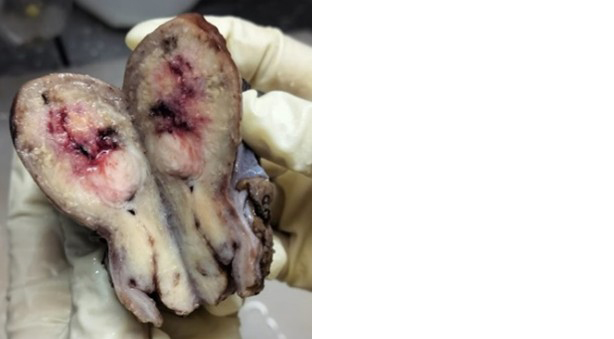
Figure 1: Cut section of the utero-cervix showing a grey-white infiltrative growth extending into the uterine corpus.

Figure 2: Cut section of the right fallopian tube showing a grey-white tumour spread compressing the lumen.
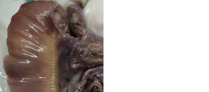
Figure 3: Cut section of the left fallopian tube showing a grey-white tumour spread compressing the lumen.
Microscopic examination revealed invasive squamous cell carcinoma of the cervix composed of neoplastic squamous cells infiltrating the cervical stroma (Figure 4). The tumour cells exhibited hyperchromatic to vesicular nuclei, prominent nucleoli, and moderate eosinophilic cytoplasm. The neoplasm extended proximally into the endometrium and myometrium in a characteristic carpet-like pattern (Figure 5). Parametrium is not involved. Both fallopian tubes showed replacement of the tubal mucosa by tumour cells. Additionally, metastatic deposits were identified in the periadnexal tissue (Figure 6). No significant tumour involvement was noted in either ovary.
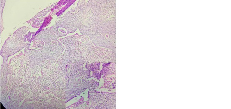
Figure 4: Microscopic image showing invasive squamous cell carcinoma of the cervix. Inset demonstrates stromal invasion.

Figure 5: Microscopic image showing carpet-like spread of cervical squamous cell carcinoma involving the endometrium and myometrium.
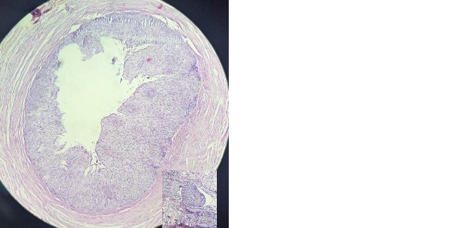
Figure 6: Microscopic image showing tumour involvement of the fallopian tube mucosa. Inset shows metastatic deposits in the periadnexal tissue.
Immunohistochemical Findings
Immunohistochemistry demonstrated p16 block positivity in the cervical tumour (Figure 7). Similar staining patterns were observed in the endometrium, myometrium (Figure 8), bilateral fallopian tubes, and periadnexal tissue (Figure 9), supporting a common cervical origin and confirming direct horizontal spread of cervical SCC to these sites.
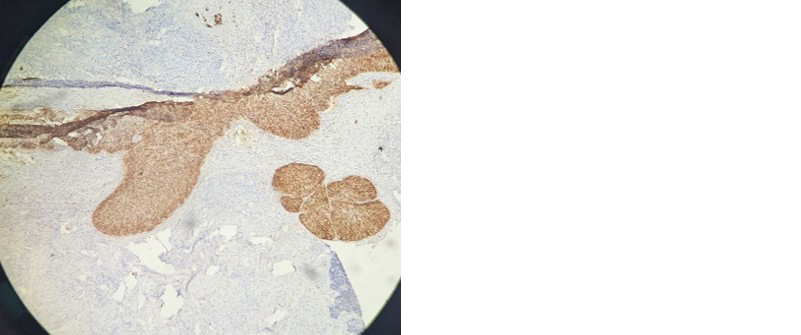
Figure 7: Immunohistochemical staining showing p16 block positivity in cervical squamous cell carcinoma.

Figure 8: Immunohistochemical staining demonstrating p16 block positivity in the uterine corpus.
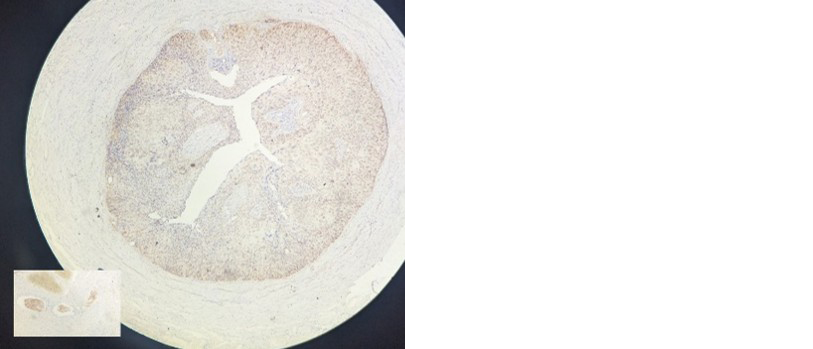
Figure 9: Immunohistochemical staining showing p16 positivity in the fallopian tube and periadnexal tissue. Inset highlights p16-positive tumour cells in periadnexal tissue.
Discussion
Our case describes an unusual pattern of cervical SCC characterized by horizontal spread to the uterine corpus, bilateral fallopian tubes, and periadnexal tissue. Cervical SCC accounts for approximately 70–80% of all cervical malignancies and usually metastasizes through lymphatic and hematogenous routes [2,3]. Superficial spread to the upper genital tract is exceptionally rare and remains poorly understood.
Endometrial involvement by cervical SCC was first described by Cullen in 1900. Subsequently, only about 50 cases have been reported in the literature, with an estimated frequency of approximately 0.7% [1]. Although the endometrium is the most common site of superficial spread, involvement of the fallopian tubes, ovaries, omentum, and transverse colon has been described only sporadically [5,6]. Martín-Vallejo et al. reviewed 54 cases of superficial spreading cervical SCC reported between 1956 and 2022, with patients ranging from 44 to 78 years of age [4]. The present case involving a 60-year-old postmenopausal woman is consistent with this demographic profile.
Several mechanisms have been proposed to explain this unusual metastatic behaviour. The horizontal theory suggests that neoplastic cells arising from a single cervical stem cell clone spread proximally along the epithelial surface, replacing the native epithelium through clonal expansion [5]. In contrast, the vertical theory postulates simultaneous malignant transformation of susceptible epithelial cells at multiple sites [8]. Persistent HPV infection, prolonged estrogen exposure, endometrial senescence, and prior cervical interventions have also been implicated as contributing factors [1,5].
Accurate differentiation from primary endometrial SCC is essential. Fluhmann proposed diagnostic criteria for primary endometrial SCC, which include the absence of coexisting endometrial adenocarcinoma, absence of primary cervical SCC, and lack of continuity between cervical and endometrial lesions [9]. In the present case, block p16 positivity in the cervix, uterine corpus, bilateral fallopian tubes, and periadnexal tissue strongly supported a common cervical origin.
HPV-associated p16 overexpression has been consistently reported in cases of superficial spread of cervical SCC. Chao et al. demonstrated HPV-p16 positivity in both endometrial and fallopian tube lesions, highlighting the pivotal role of HPV in this process [10]. Similar findings were observed in our case.
The current FIGO 2018 and AJCC Version 9 staging systems do not specifically address horizontal spread of cervical carcinoma to the uterine corpus and adnexa [11]. Consequently, there is limited guidance regarding staging, prognosis, and management of these patients. Future studies focusing on molecular mechanisms and targeted therapeutic strategies are warranted to improve clinical outcomes.
Conclusion
Cervical squamous cell carcinoma may exhibit rare but clinically significant horizontal spread to the uterine corpus and adnexal structures. Recognition of this unusual metastatic pattern is important because it can mimic primary endometrial or adnexal neoplasms and may influence clinical management. Histopathological examination supplemented by immunohistochemistry, particularly p16 staining, plays a pivotal role in establishing the diagnosis and confirming the cervical origin of the tumour. Clinicians and pathologists should maintain a high index of suspicion in postmenopausal women presenting with vaginal bleeding, cervical stenosis, or pyometra. Further studies are required to elucidate the underlying mechanisms, prognostic implications, and optimal management strategies for this uncommon pattern of tumour spread.
Conflicts of interest
Authors declare no conflicts of interest.
References
[1] Shu XY, Dai Z, Zhang S, Yang HX, Bi H. Endometrial squamous cell carcinoma originating from the cervix: A case report. World J Clin Cases. 2022; 10:8782–8787.
[2] Reyes C, Murali R, Park KJ. Secondary involvement of the adnexa and uterine corpus by carcinomas of the uterine cervix: a detailed morphologic description. Int J Gynecol Pathol. 2015; 34:551–563.
[3] Rangankar V, Bokil S, Deshmukh S, Bokil SS. Superficial spread of nonkeratinizing squamous cell carcinoma of the cervix to the endometrium: A case report. Cureus. 2024; 16.
[4] Martin–Vallejo J, Laforga JB, Molina–Bellido P, Clemente–Perez PA. Superficial spreading cervical squamous cell carcinoma in situ involving the endometrium: a case report and review of the literature. J Med Case Rep. 2022; 16:196.
[5] Nakajima T, Hatta H, Nishida T, Minamisaka T, Miwa S, et al. Superficial spread of cervical squamous cell carcinoma to the upper genital tract and dissemination to the omentum. Pathol Int. 2019; 69:119–121.
[6] Kushima M, Fujii H, Murakami K, Ota H, Matsumoto T, et al. Simultaneous squamous cell carcinomas of the uterine cervix and upper genital tract: loss of heterozygosity analysis demonstrates clonal neoplasms of cervical origin. Int J Gynecol Pathol. 2001; 20:353–358.
[7] Dokic M, Milenkovic S, Jovanovic L, Milosevic B, Andric L, et al. Curious case of superficial spreading cervical squamocellular carcinoma with adnexal involvement. Medicina (Kaunas). 2022; 58:1655.
[8] Sood N, Sinha KS. Superficial spreading squamous cell carcinoma endometrium andicthyosis uteri with CINIII with p16 expression: Report of 2 unusual cases. J. Krishna Inst. Med. Sci. Univ. 2017; 6:126–131.
[9] Fluhmann CF. The histogenesis of squamous cell metaplasia of the cervix and endometrium. Surg Gynecol Obstet. 1953; 97:45–58.
[10] Chao A, Wang AM, Wang TH, Wu TI, Chao AS. An atypical and fatal case of pyometra accompanied by the superficial spread of squamous cell carcinoma of the endometrium and the fallopian tubes. Taiwan J Obstet Gynecol. 2013; 52:440–442.
[11] Olawaiye AB, Baker TP, Washington MK, Mutch DG. The new (Version 9) American Joint Committee on Cancer tumor, node, metastasis staging for cervical cancer. CA Cancer J Clin. 2021; 71:287–298.