Full Text
Introduction
Fractures of the distal humerus with inter-condylar extension in adults present a great challenge to even the most experienced surgeon due to the complex anatomical configuration of elbow joint, small and multiple fractured fragments, osteopenia of articulating surfaces and limited amount of subchondral bone [1, 2]. The functional outcome of these type of fractures is related to the ability to restore the normal elbow anatomy and to achieve good functional range of motion at elbow joint and early movement. The various methods described in literature to fix such type of fractures are Kirschner wires (K-wires), screw fixation, sole plate but the stability provided by these methods is not good enough to start early mobilization of the elbow joint [3-6]. Recently, with the advancement of science and technology which led to the development of anatomical plates which are pre-contoured, most of the orthopaedic surgeons fix these fractures by more than 1 screw in intercondylar region, but none of these screws has a purchase on opposite cortex, thereby making this fixation not very stable and rigid which can drastically affect the fracture and cause its non-union.
In this study, we have tried to use a simple, inexpensive, easier and reliable method of fixing these fractures of intercondylar region of distal humerus and maintaining the normal anatomy by fixing all the fractured fragments by using K-wires and SS wires i.e., by using double tension band wiring technique in one group of 20 patients. In this technique, the humerus condyles are first reduced with cannulated cancellous screws and which are later fixed to humerus shaft with double tension bands and these are tied across a screw inserted in the midline of humerus shaft on posterior surface. The principle used in these patients was that of Tension Band Wiring to achieve rigid fixation of both the columns of humerus. This principle applies to conversion of tensile forces at the fracture site to compressive forces on the convex side of bone [3] thus enabling contact at the fracture site. This technique required minimal use of implant, is inexpensive, simple and less operative time thus leading to less blood loss and less rate of complications over the locking plates.
In another group of 30 patients, we have used method of internal fixation i.e. locking plates of distal humerus. In this technique, we first reduced the fractured fragments anatomically and used cannulated cancellous screws to fix inter-condylar fracture. Then we provisionally fixed the fracture with k-wires. Later, locking plates were used on medial and lateral column of distal humerus in two configurations in which these plates can be used are parallel and orthogonal plating. By this method we achieved rigid fixation of fractured fragments and started early mobilization at elbow joint.
The study aimed to observe the effectiveness of “double tension band wiring” method for the remedy of distal humerus fractures with inter-condylar extension and compare the effects with studies related to its treatment with locking plates.
Materials and methods
The study was carried out from April 2017 to May 2023 at Kalpana Chawla Government Medical College, Haryana, 50 patients of supracondylar fracture of humerus with inter-condylar extension were operated using both these techniques. It was a retrospective study with collection of data from patients in OPD after every follow up visit. The patients were enrolled serially as per operation record and data collection with x-ray and other details were taken during follow up period. Both the groups of plating versus tension BAND wiring were comparable by their demographic situation as all were localized in Haryana as well as by similar fracture patterns. All adult patients having C1 type intercondylar with fracture of the distal humerus were selected. Standard anterior-posterior and lateral radiographs were obtained in the radiology department and all fractures were classified according to Jupiter classification [4] of distal humeral fractures (Table 1). All the selected patients were divided in two groups. In the first group, we included 20 patients which were operated with double tension band wiring technique. In other group we included 30 patients and operated with locking plates. There were 15 high T fractures, 30 low T fractures and 5 Y fractures. According to AO classification, all were C1 fractures [5]. The mean age of presentation of patients was 55 years (33-68 years). There were 41 males and 9 female among the 50 patients.
Table 1: Jupiter classification of distal humerus fractures.
|
High-T
|
Transverse fracture proximal to or at upper olecranon fossa
|
|
Low-T
|
Transverse fracture just proximal to trochlea
|
|
Y
|
Oblique fracture line through both columns with distal vertical fracture line
|
|
H
|
Trochlea is a free fragment (risk of AVN)
|
|
Medial lambda
|
Proximal fracture line exists medially
|
|
Lateral lambda
|
Proximal fracture line exists laterally
|
|
Multiplane T
|
T type with additional fracture in coronal plane
|
All patients were operated in lateral position under regional block or general anaesthesia. Upper limb pneumatic tourniquet was used and was inflated just before making the skin incision. We used posterior approach to elbow joint in which midline posterior incision given and soft tissue dissection done. Ulnar nerve was identified and retracted to avoid injury. Then Chevron osteotomy for olecranon was done. Fracture site exposed and inter-condylar fragments of fracture were first reduced and held with AO reduction clamp or K-wire as anatomical as possible. Then we passed a guide wire for cannulated cancellous screw from medial side to lateral aspect or vice versa in intercondylar area crossing fracture perpendicularly. A cannulated cancellous drill bit was used to drill over the guide wire and a proper sized 4mm cannulated cancellous screw was inserted over the guide wire to secure the fragments in anatomical position.
Now in first group, the condylar block was attached to the humeral shaft using double tension band. Firstly, after reducing the condylar block to the humeral shaft, two K-wires were passed from the lateral condyle across the fracture to engage the medial cortex of humeral shaft and likewise two K-wires were passed from the medial condyle across the fracture to engage the lateral cortex of the humeral shaft. Now a 4.5 mm cortical screw with a washer was inserted in the midline of humeral shaft just above the fracture at the level of K-wire tips. An 18 gauge stainless steel wire was passed beneath the screw head and across the medial K-wire tips distally in figure of 8 manner and tightened using double loop. Similarly a tension band was applied laterally. Stability of the construct was checked under vision and olecranon osteotomy was closed using tension band wiring (Figure 1). Again the stability of the whole construct was checked by moving the elbow through full range of motion.
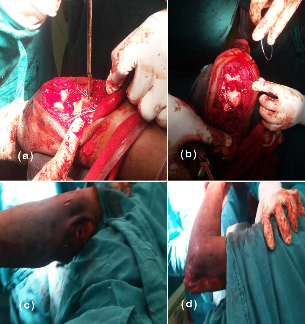
Figure 1: (a) Tension band wiring seen with fixation in both condyles of humerus, (b) SS wire being applied, (c) Range of motion being checked after Tension band wiring with elbow in flexion and extension, (d) Range of motion was being checked after Tension band wiring with elbow in extension.
In another group of 30 patients, we have used method of internal fixation i.e., locking plates of distal humerus. In this technique, we first reduced the fractured fragments anatomically and used cannulated cancellous screws to fix inter-condylar fracture. Then we provisionally fixed the fracture with k-wires. Later, locking plates were used on medial and lateral column of distal humerus in 90-90 position. By this method we achieved rigid fixation of fractured fragments.
Ulnar nerve was transposed anteriorly when needed. Tourniquet was deflated and haemostasis achieved. Wound was closed in layers over a suction drain. Drain was removed on second postoperative day and gentle active range of motion exercises was started. Patients were discharged from the hospital after suture removal around 11 or 12 days and followed up in OPD at 3 weeks, 4 weeks, 6 weeks and then monthly up to one year (Figures 2 to 9). Final result was graded according to criteria laid by Jupiter et al., (Table 2). General patient information has been provided in (Tables 3 and 4).
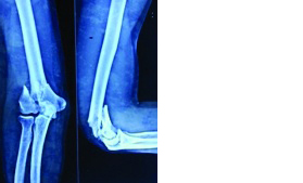
Figure 2: (a) Anterior posterior x-ray view of elbow shows both lateral and medial condyle fracture, (b) Lateral view x-ray shows comminuted distal humerus fracture.
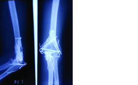
Figure 3: Post-operative radiograph at 10 weeks follow up (a) Lateral view x-ray shows comminuted distal humerus fracture under process of union, (b) Anterior posterior x-ray view of elbow shows both lateral and medial condyle fracture under process of union.
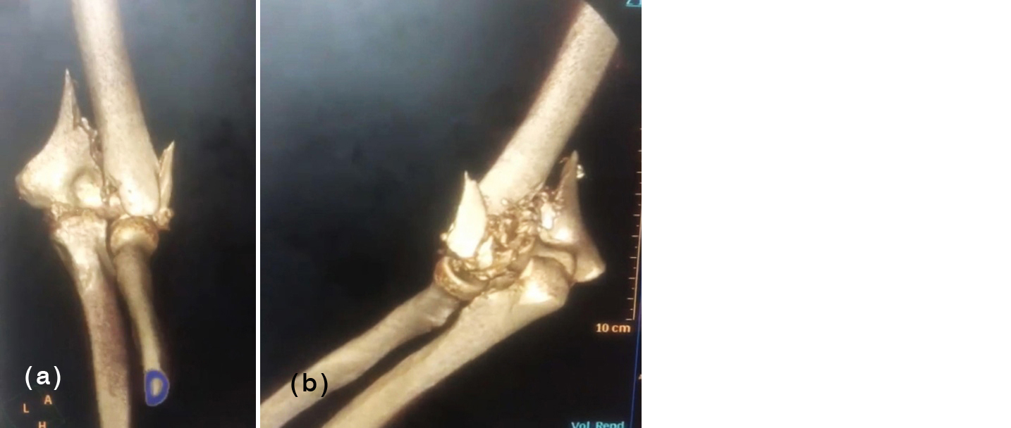
Figure 4: (a) Anterior posterior 3D CT scan view of elbow shows both lateral and medial condyle fracture, (b) Lateral view 3D CT scan shows comminuted distal humerus fracture.
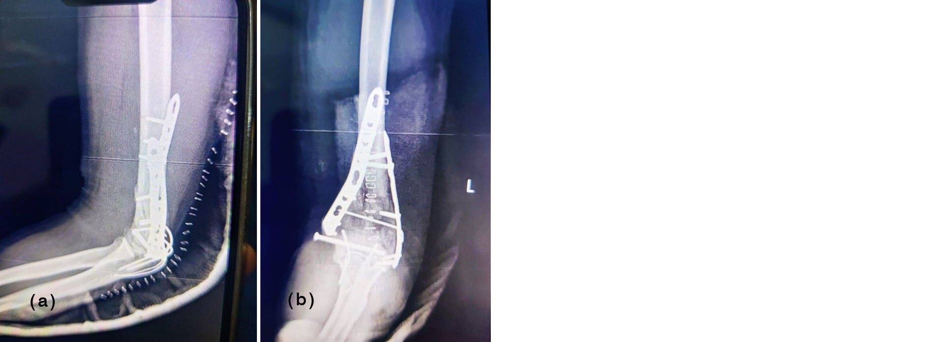
Figure 5: ORIF with bi-columnar plating (a) Lateral view x-ray depicts fracture uniting with implants in situ, (b) Anterior posterior view x-ray depicts fracture uniting with implants in situ.
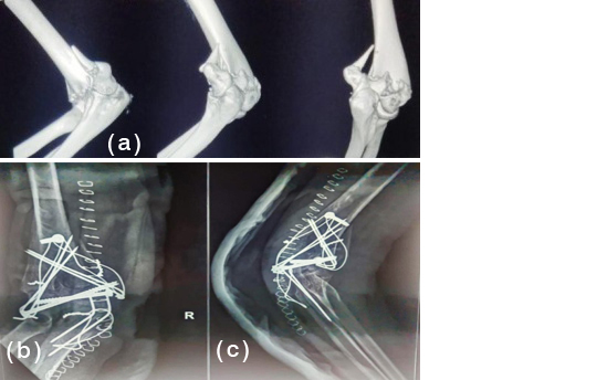
Figure 6: Another case (a) Preoperative 3D CT scan view of elbow shows both lateral and medial condyle fracture, (b) x-ray shows comminuted distal humerus fracture under process of union in anterior posterior view, (c) x-ray shows comminuted distal humerus fracture under process of union in lateral view.
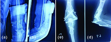
Figure 7: (a) Anterior posterior x-ray view of elbow shows both lateral and medial condyle fracture, (b) Lateral view x-ray shows comminuted distal humerus fracture, (c) Tension band wiring seen with fixation in both condyles of humerus in AP view, (d) Tension band wiring seen with fixation in both condyles of humerus in lateral view.
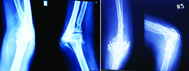
Figure 8: (a) Anterior posterior x-ray view of elbow shows both lateral and medial condyle fracture, (b) Lateral view x-ray shows comminuted distal humerus fracture, (c) Tension band wiring seen with fixation in both condyles of humerus in AP view, (d) Tension band wiring seen with fixation in both condyles of humerus in lateral view.

Figure 9: Patient with full range of motion (a) complete pain free extension, (b) complete pain free flexion.
Table 2: Grading criteria by Jupiter et al., [4].
|
Result
|
Loss of extension
|
Flexion
|
Pain
|
Disability
|
|
Excellent
|
<15°
|
>130°
|
None
|
None
|
|
Good
|
<30°
|
>120°
|
Slight
|
Minimal
|
|
Fair
|
<40°
|
>90°
|
With activity
|
Moderate
|
|
Poor
|
<40°
|
>90°
|
Variable
|
Severe
|
Table 3: Group 1- General patient information sheet.
|
Age
|
Sex
|
Jupiter class
|
Tourniquet time (in minutes)
|
Radiological union
(In weeks)
|
Extension (in degrees)
|
Flexion (in degrees)
|
Results
|
|
32
|
Male
|
Y
|
60
|
8
|
-10
|
130
|
Excellent
|
|
63
|
Female
|
High T
|
50
|
12
|
-20
|
120
|
Good
|
|
65
|
Male
|
Low T
|
120
|
14
|
-25
|
120
|
Good
|
|
58
|
Female
|
Low T
|
80
|
10
|
-15
|
120
|
Good
|
|
65
|
Male
|
High T
|
50
|
12
|
-20
|
120
|
Good
|
|
70
|
Female
|
Low T
|
60
|
10
|
-30
|
100
|
Fair
|
|
55
|
Male
|
Low T
|
110
|
12
|
-10
|
125
|
Good
|
|
61
|
Male
|
Low T
|
90
|
14
|
-20
|
110
|
Fair
|
|
50
|
Male
|
High T
|
60
|
10
|
-15
|
130
|
Excellent
|
|
45
|
Male
|
Low T
|
70
|
9
|
-10
|
120
|
Good
|
|
55
|
Female
|
Low T
|
60
|
10
|
-15
|
100
|
Fair
|
|
56
|
Male
|
High T
|
80
|
12
|
-10
|
120
|
Good
|
|
68
|
Male
|
Low T
|
60
|
8
|
10
|
130
|
Excellent
|
|
60
|
Male
|
Low T
|
60
|
12
|
15
|
110
|
Fair
|
|
56
|
Male
|
Low T
|
50
|
10
|
10
|
130
|
Excellent
|
|
52
|
Male
|
Low T
|
70
|
14
|
10
|
120
|
Good
|
|
58
|
Male
|
High T
|
50
|
10
|
10
|
120
|
Good
|
|
55
|
Male
|
Y
|
60
|
9
|
10
|
120
|
Good
|
|
50
|
Male
|
Low T
|
80
|
10
|
15
|
120
|
Good
|
|
50
|
Male
|
Low T
|
60
|
10
|
15
|
110
|
Fair
|
Table 4: Group 2- General patient information sheet.
|
Age
|
Sex
|
Jupiter class
|
Tourniquet time (in minutes)
|
Radiological union
(In xeeks)
|
Extension (in degrees)
|
Flexion (in degrees)
|
Results
|
|
42
|
Male
|
Y
|
60
|
8
|
-10
|
130
|
Excellent
|
|
53
|
Female
|
High T
|
50
|
12
|
-20
|
120
|
Good
|
|
55
|
Male
|
Low T
|
120
|
14
|
-25
|
120
|
Good
|
|
54
|
Female
|
Low T
|
80
|
10
|
-15
|
120
|
Good
|
|
75
|
Male
|
High T
|
50
|
12
|
-20
|
120
|
Good
|
|
60
|
Male
|
Low T
|
60
|
10
|
-30
|
100
|
Fair
|
|
45
|
Male
|
Low T
|
110
|
12
|
-10
|
125
|
Good
|
|
51
|
Female
|
Low T
|
90
|
14
|
-20
|
110
|
Fair
|
|
60
|
Male
|
High T
|
60
|
10
|
-15
|
130
|
Excellent
|
|
55
|
Male
|
Low T
|
70
|
9
|
-10
|
120
|
Good
|
|
65
|
Female
|
Low T
|
60
|
10
|
-15
|
100
|
Fair
|
|
66
|
Male
|
High T
|
80
|
12
|
-10
|
120
|
Good
|
|
48
|
Male
|
Low T
|
60
|
8
|
10
|
130
|
Excellent
|
|
50
|
Male
|
Low T
|
60
|
12
|
15
|
110
|
Fair
|
|
58
|
Male
|
Low T
|
50
|
10
|
10
|
130
|
Excellent
|
|
51
|
Male
|
Low T
|
70
|
14
|
10
|
120
|
Good
|
|
59
|
Male
|
High T
|
50
|
10
|
10
|
120
|
Good
|
|
51
|
Male
|
Y
|
60
|
9
|
10
|
120
|
Good
|
|
40
|
Male
|
Low T
|
80
|
10
|
15
|
120
|
Good
|
|
46
|
Male
|
Low T
|
60
|
10
|
15
|
110
|
Fair
|
|
65
|
Female
|
Low T
|
120
|
14
|
-25
|
120
|
Good
|
|
58
|
Male
|
Low T
|
80
|
10
|
-15
|
120
|
Good
|
|
65
|
Male
|
High T
|
50
|
12
|
-20
|
120
|
Good
|
|
70
|
Female
|
Low T
|
60
|
10
|
-30
|
100
|
Fair
|
|
55
|
Male
|
Low T
|
110
|
12
|
-10
|
125
|
Good
|
|
61
|
Male
|
Low T
|
90
|
14
|
-20
|
110
|
Fair
|
|
50
|
Male
|
High T
|
60
|
10
|
-15
|
130
|
Excellent
|
|
45
|
Male
|
Low T
|
70
|
9
|
-10
|
120
|
Good
|
|
55
|
Female
|
Low T
|
60
|
10
|
-15
|
100
|
Fair
|
|
56
|
Male
|
High T
|
80
|
12
|
-10
|
120
|
Good
|
Results
Out of 50 cases treated with these 2 methods of internal fixation, rigid fixation and union was achieved in each and every patient. The mean duration of use of Tourniquet during surgery was 65 minutes with minimum of 54 minutes and maximum of 110 minutes. Complete radiological union of the fracture was seen at a mean duration of 11 weeks (Figure 10) after the surgery (9-15 weeks).
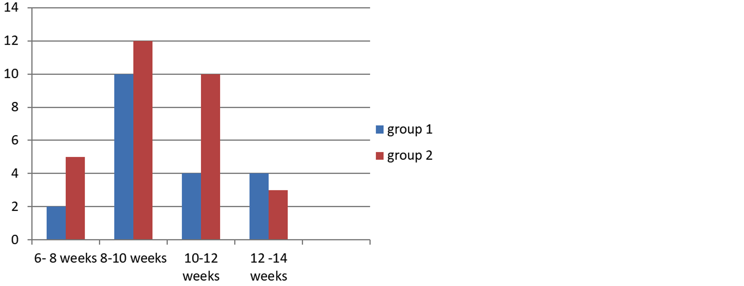
Figure 10: Histogram showing radiological union in both groups in weeks.
Average range of motion was 103 degrees (Figures 11 and 12) with maximum range of motion of 110 degrees (20-130 degrees) and minimum of 65 degrees (25-90 degrees). Good results were obtained in both the groups in our study. The loss of range of motion was consistent with the age of the patient and compliance of the patient with physical therapy. Three patients had ulnar nerve neuropraxia in the postoperative period which corrected over a period of 5 weeks via conservative approach. Two patients had palpable k-wire tips beneath the skin which were removed after radiological union. Overall patients had minimal symptoms related to hardware.

Figure 11: group 1- elbow range of motion.

Figure 12: group 2- elbow range of motion.
Discussion
Fixation of intra-articular distal humerus fractures represents a challenging reconstructive problem for orthopaedic surgeons due to complex anatomy of the distal humerus and difficulty in exposing the fracture which is further compounded by comminution and osteopenia of articulating surfaces. The main goal in such type of fractures is anatomical reduction, rigid fixation and early mobilisation required to prevent complications [7]. We have compared two such methods here which allow good compression at fracture site, rigid fixation utilising minimal hardware. The method of double tension band wiring of distal humerus has the advantage of early mobilization which can be attributed to the rigid fixation provided by the tension band. The amount of periosteal stripping & soft tissue damage is significantly less than plate fixation. It is a simple procedure and the duration of the surgeries in our series was around 90 minutes. It is inexpensive when compared to the costs of plates. Tourniquet time can be minimised with this technique preventing tourniquet related complications. Because of the rigidity and stability achieved in this technique and because the tension band wiring technique acts in dynamic mode when muscles contract, we can mobilise the patient very early. In fractures in osteoporotic bone where large implants can have shattering effect and screw may loosen out, this technique gives good hold without fear of loosening with minimal implants.
Double tension band wiring has been used in past with encouraging results. In a study of 10 patients, Houben et al reported comparable results, where 5 patients were treated with double tension band and 5 were treated with double plates [8]. Zhao et al reported 83% excellent or good results in their study of 24 patients treated with double tension band wiring [9]. This method can also be used in combination with other methods of fixation, as reported by Allende et al [10], where they concluded that the method is good alternative in osteoporotic settings when combined with other methods.
Presently, there is increasing trend towards the use of locking plates in intercondylar fractures of distal humerus. The very design of the locked plates is for osteoporotic fractures [11–13]. These plates can be used in 2 different configurations: parallel plaing or orthogonal plating. There is no significant difference with regards to clinical and functional outcomes in using the plates in different configurations. The decision to use parallel plating or orthogonal plating depends on surgeon’s preference and bone quality.
There are some disadvantages of these plates like the extensive soft tissue damage, technical expertise required, increased operative time and issues with contouring in some patients. The advantage of these plates, when compared with double tension band osteosynthesis lies in their biomechanical strength, as reported by Dogramacai et al [14].
Success in treating these fractures starts with pre-operative understanding of the normal anatomy of distal humerus and the fracture pattern. Intraoperatively, obtaining an anatomical reduction of the articular surface with a stable hardware construct, which will allow early range of motion while minimizing complications, will surely result in favourable outcomes.
We have not studied the biomechanical strength of the tension band but since we were able to start early mobilization, all fractures united & there was no breakage of any wires; it can be postulated that this method has enough biomechanical stability to be used in clinical practice. Dubey et al described a modification and did transosseous fixation of intercondylar fracture of lower end humerus by tension band wiring technique, and reported excellent results [15].
Our study shows that in inter-condylar fractures in adults, double tension band fixation can be a safe, reliable, easy and inexpensive technique for the management of these fractures and should remain an option to be considered when planning the stabilisation of these fractures especially the AO type C1 fractures. Our results are comparable to other studies done using locked plates.
Limitations of the study: The above study although was done in a tertiary care medical college has limited patients; multi–centre trial with more patients gives precise results. Admittedly, the present study is based on a limited number of cases and is inadequate to provide conclusive data
Conclusion
Double tension band is a reliable, more biological, less surgically demanding and cost effective method of fixation of intercondylar fractures of humerus. Rigid fixation and hence, early mobilisation can be achieved with this method. This method avoids extensive soft tissue stripping and bulky hardware. Though locked plates are considered as gold standard in the treatment of fractures of the distal humerus, double tension band technique should be considered as an option while planning fixing these fractures especially AO type C1 fractures. The main limitations of this series are having a small number of patients and no experience with communited AO type C2 and C3 fractures.
Conflicts of interest
Authors declare no conflicts of interest.
References
[1] Cannada L, Loeffler B, Zadnik MB, Eglseder AW. Treatment of high-energy supracondylar/intercondylar fractures of the distal humerus. J Surg Orthop Adv. 2021; 20:230–35.
[2] Schmidt-Horlohé K, Wilde P, Bonk A, Becker L, Hoffmann R. One-third tubular-hook-plate osteosynthesis for olecranon osteotomies in distal humerus type-C fractures: a preliminary report of results and complications. Injury. 2020; 43:295–300.
[3] Pauwels F. Biomechanics of the locomotor apparatus. Berlin Heidelberg New York: Springer-Verlag; 2019.
[4] Jupiter JB, Mehne DK. Fractures of the distal humerus. Orthopedics. 2018; 15:825–33.
[5] Ruedi T, Murphy WM. AO principles of fracture management. Vol. 1. Thieme: Stuttgart-New York; 2018.
[6] Jupiter JB, Noff U, Hoizach P, Allgower M. Intercondylar fractures of the humerus: An operative approach. J Bone Joint Surg (Am) 2018; 67:226–39.
[7] Gupta R, Khanchandani P. Intercondylar fractures of the distal humerus in adults: a critical analysis of 55 cases. Injury. 2018; 33:511–515.
[8] Houben PFJ, Bongers KJ, Wildenberg FAJM. Double band osteosynthesis in supra- and transcondylar humeral fractures. Injury. 2017; 25:305–309.
[9] Zhao J, Wang X, Zhang Q. Surgical treatment of comminuted intra-articular fractures of the distal humerus with double tension band osteosynthesis. Orthopaedics. 2016; 23:449–452.
[10] Allende CA, Allende BT, Allende BL, Gonzalez GBI. Intercondylar distal humerus fractures—surgical treatment and results. Chir Main. 2015; 23:85–95.
[11] Gupta RK, Gupta V, Marak DR. Locking plates in distal humerus fractures: Study of 43 patients. Chinese J Traumatol. 2013; 16:207–211.
[12] Reising K, Hauschild O, Strohm PC, Suedkamp NP. Stabilization of articular fractures of the distal humerus: early experience with a novel perpendicular plate system. Injury. 2009; 40:611–617.
[13] Greiner S, Haas NP, Bail HJ. Outcome after open reduction and angular stable internal fixation for supra-intercondylar fractures of the distal humerus: preliminary results with the LCP distal humerus system. Arch Orthop Trauma Surg. 2008; 128:723–29.
[14] Dogramaci Y, Esen E, Kürklü M, Kirici Y, Atahan AO, et al. Double plate osteosynthesis provides better biomechanical stabilization than double tension band technique in distal humerus fractures. Eklem Hastalik Cerrahisi. 2008; 21:44–49.
[15] Dubey R, Sadigale V, Parmar J. Transosseous fixation of intercondylar fracture of lower end humerus by tension band wiring technique. Int J Med Sci Public Health. 2007; 3:305–307.