Full Text
Introduction
Total knee arthroplasty (TKA) is an established treatment for end-stage knee osteoarthritis and is effective in relieving pain, correcting deformity, and improving function [1]. The increasing prevalence of osteoarthritis and the growing number of older adults have contributed to a continuing rise in demand for primary TKA.
Obesity is an important risk factor for the development and progression of knee osteoarthritis and is increasingly encountered among patients undergoing TKA [2, 3]. Increased body mass places greater mechanical demands on the knee prosthesis and may increase the risk of complications, including aseptic tibial loosening, infection, and revision surgery [4, 5]. In particular, a higher BMI has been associated with an increased risk of aseptic tibial failure even when postoperative alignment is satisfactory [4].
These concerns have led to interest in tibial stem extensions as a means of improving fixation and distributing load more effectively from the tibial baseplate toward the metaphyseal or diaphyseal bone. However, stemmed components may increase implant cost and technical complexity and may have disadvantages during revision surgery. Evidence comparing stemmed and non-stemmed tibial components in obese patients remains mixed, with some studies reporting possible advantages and others demonstrating comparable clinical outcomes [6–9].
Assessment of postoperative pain and functional recovery using validated outcome measures is important when comparing implant designs. The present study was therefore undertaken to compare postoperative pain, functional outcome, radiological alignment, and postoperative complications following primary TKA using stemmed versus non-stemmed tibial components in obese patients.
The objective of this study was to determine whether the use of a tibial stem extension provides superior postoperative pain relief and functional recovery compared with a conventional non-stemmed tibial component in obese patients undergoing primary TKA.
Materials and methods
This retrospective comparative study was conducted in the Department of Orthopaedics at Krishna Institute of Medical Sciences (KIMS), Secunderabad, Hyderabad. Hospital records of patients who underwent primary TKA and had completed a minimum follow-up of 1 year were retrospectively reviewed from February 2025 to January 2026. The study has been approved by institutional ethics committee.
A total of 20 patients who underwent primary TKA were included. Ten patients had received a stemmed tibial component (Group A), and 10 had received a non-stemmed tibial component (Group B).
Inclusion criteria: Patients were eligible if they had primary osteoarthritis of the knee, a BMI >30 and <40 kg/m², an age between 40 and 70 years, a varus deformity of <15°, and had undergone primary TKA using either a stemmed or non-stemmed tibial component.
Exclusion criteria: Patients were excluded if they had valgus deformity, a fixed flexion deformity >10°, previous surgery on the affected knee, revision TKA, poor bone quality on radiographic or intraoperative assessment, incomplete medical records, or inadequate follow-up. Poor bone quality was identified using the Reverse Dorr radiographic assessment and/or documented intraoperative findings such as sparse or porous cancellous bone, poor implant purchase, bone crumbling during preparation, or the need for augmentation.
Bone-quality assessment
The study records state that the Reverse Dorr radiographic method was used as a preoperative screening tool for bone quality. Patients with a Reverse Dorr ratio <1.37 were considered to have radiographic evidence suggestive of poor bone quality and were excluded. Because this is a retrospective screening method rather than a definitive diagnosis of osteoporosis, the findings should be interpreted as a study-specific exclusion criterion rather than as a substitute for bone mineral density measurement [10].
Data collection
Demographic and clinical information was collected retrospectively from hospital medical records, operative notes, radiographs, and follow-up records. Variables included age, sex, BMI, type of tibial component, postoperative pain score, functional KSS, radiological β angle, and postoperative complications.
Outcome measures
The primary clinical outcomes were postoperative pain and functional outcome. Pain was assessed using the VAS, scored from 0 (no pain) to 10 (worst possible pain). Functional outcome was assessed using the Functional Knee Society Score, with higher scores indicating better knee function. The latest available postoperative VAS and KSS documented during routine follow-up were used for analysis. Radiological outcome was assessed using the tibial component β angle, and postoperative complications documented in the medical records were recorded.
Statistical analysis
Data were entered into Microsoft Excel and analysed using IBM SPSS Statistics. Continuous variables were summarized as mean ± standard deviation, and categorical variables were summarized as frequencies and percentages. Between-group comparisons were performed using the independent-samples Student’s t-test for continuous variables and the chi-square test or Fisher’s exact test for categorical variables, as appropriate. A p-value <0.05 was considered statistically significant.
Results
A total of 20 patients were included in the analysis, with 10 patients in the stemmed group and 10 in the non-stemmed group. Baseline demographic characteristics are presented in Table 1. The mean age was 64.4 ± 5.4 years in the stemmed group and 60.3 ± 8.8 years in the non-stemmed group (p=0.228), while the mean BMI was 31.90 ± 2.15 kg/m² and 30.75 ± 0.87 kg/m², respectively (p=0.146). The sex distribution was also comparable between groups (Table 1).
Table 1: Baseline demographic characteristics.
|
Variable
|
Stemmed (n=10)
|
Non-stemmed (n=10)
|
p-value
|
|
Age (years)
|
64.4 ± 5.4
|
60.3 ± 8.8
|
0.228
|
|
BMI (kg/m²)
|
31.90 ± 2.15
|
30.75 ± 0.87
|
0.146
|
|
Male
|
2 (20%)
|
4 (40%)
|
0.626
|
|
Female
|
8 (80%)
|
6 (60%)
|
|
Postoperative pain outcomes are summarized in Table 2. The mean VAS score was 2.10 ± 1.29 in patients with stemmed tibial components and 2.50 ± 0.97 in those with non-stemmed components. Although the mean score was numerically lower in the stemmed group, the between-group difference was not statistically significant (p=0.444), indicating comparable postoperative pain outcomes.
Table 2: Comparison of postoperative pain.
|
Variable
|
Stemmed
|
Non-stemmed
|
p-value
|
|
VAS score
|
2.10 ± 1.29
|
2.50 ± 0.97
|
0.444
|
Functional outcomes are presented in Table 3. The mean Functional KSS was 71.0 ± 18.53 in the stemmed group and 69.0 ± 9.94 in the non-stemmed group. The difference was not statistically significant (p=0.768), suggesting that both implant designs provided comparable short-term functional recovery.
Table 3: Comparison of functional outcome.
|
Variable
|
Stemmed
|
Non-stemmed
|
p-value
|
|
Functional KSS
|
71.0 ± 18.53
|
69.0 ± 9.94
|
0.768
|
Radiological alignment and postoperative complications are summarized in Table 4. The mean tibial component β angle was 89.41° ± 1.34° in the stemmed group and 90.23° ± 1.34° in the non-stemmed group, with no statistically significant difference (p=0.189). One postoperative complication was documented in the stemmed group compared with none in the non-stemmed group; this difference was not statistically significant (Fisher’s exact test, p=1.000).
Table 4: Comparison of radiological outcome and complications.
|
Variable
|
Stemmed
|
Non-stemmed
|
p-value
|
|
β angle (°)
|
89.41 ± 1.34
|
90.23 ± 1.34
|
0.189
|
|
Complications
|
1 (10%)
|
0
|
1.000*
|
*Fisher’s exact test.
Figure 1 shows the postoperative standing radiograph of a 69-year-old woman with a BMI of 30.1 kg/m² at 13 months after primary TKA with a non-stemmed tibial component. The image demonstrates the postoperative appearance and alignment of the non-stemmed tibial component at follow-up.
Figure 2 shows the postoperative radiograph of a 57-year-old woman with a BMI of 33.27 kg/m² at 1 year after primary TKA with a stemmed tibial component. The image demonstrates the postoperative appearance of the stemmed tibial component at follow-up.
Figure 3 demonstrates measurement of the tibial component β angle in a patient with a stemmed tibial implant at 1-year follow-up. The β angle shown in the figure is 90.7°, illustrating the radiographic method used for assessment of tibial component alignment.
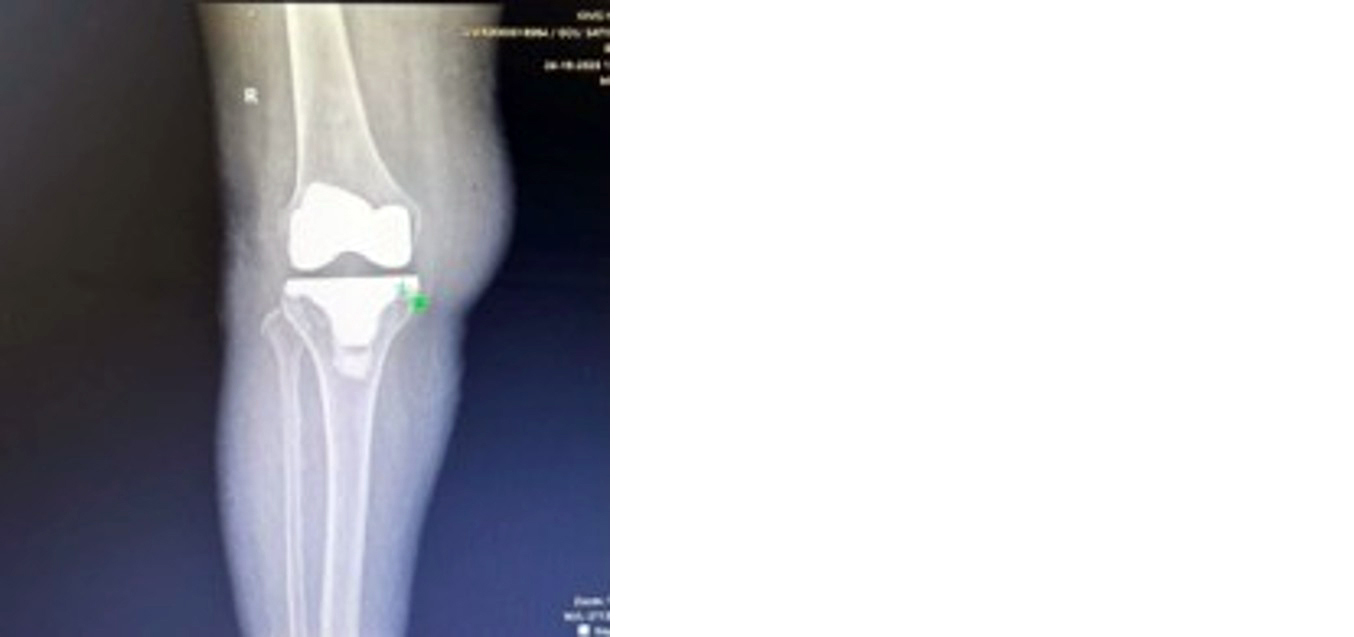
Figure 1: Postoperative radiograph of a 69-year-old woman with a BMI of 30.1 kg/m² at 13 months after primary TKA with a non-stemmed tibial component.
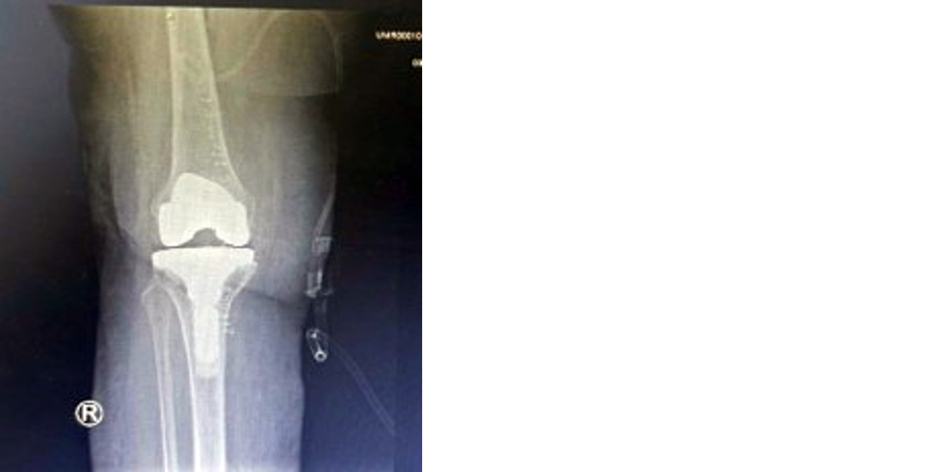
Figure 2: Postoperative radiograph of a 57-year-old woman with a BMI of 33.27 kg/m² at 1 year after primary TKA with a stemmed tibial component.
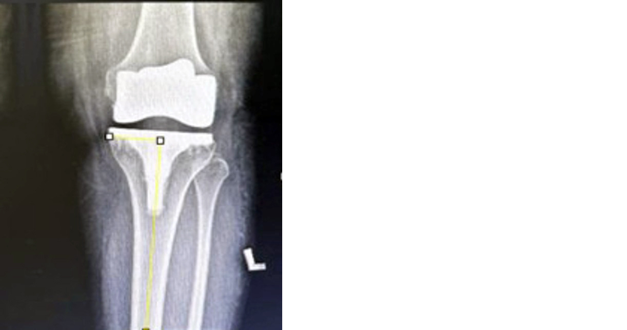
Figure 3: Radiographic measurement of the tibial component β angle in a patient with a stemmed tibial implant at 1-year follow-up; the measured β angle is 90.7°.
Discussion
The present retrospective comparative study evaluated whether stemmed tibial components provide superior short-term clinical and radiological outcomes compared with non-stemmed tibial components in obese patients undergoing primary TKA. In this cohort of 20 patients, postoperative pain, functional outcome, tibial component alignment, and complication rates were comparable between the two groups.
Obesity is associated with increased mechanical loading across the knee and has been linked to a higher risk of complications and revision after TKA [11-13]. Berend et al. reported that a higher BMI was associated with medial tibial bone collapse and identified BMI >33.7 kg/m² as one of the factors associated with tibial component failure [11]. Studies of TKA outcomes in obese patients have also reported increased risks of complications and revision, supporting the importance of careful implant selection in this population [12,13]. However, increased mechanical risk does not necessarily mean that every obese patient requires a tibial stem extension.
In the present study, postoperative VAS scores were slightly lower in the stemmed group, but the difference was not statistically significant (Table 2). Similarly, Functional KSS did not differ significantly between the groups (Table 3). These findings suggest that, within the limitations of this small retrospective cohort, the addition of a tibial stem did not confer a demonstrable short-term functional or pain-related advantage.
These findings are broadly consistent with previous studies evaluating the use of tibial stems in primary TKA [14-16]. Studies examining tibial stem extensions have reported that their use may provide additional fixation and improve load distribution in selected patients, particularly those with obesity or other factors associated with increased mechanical demand [14-16]. However, the available evidence does not establish that all obese patients undergoing primary TKA require a stemmed tibial component. Therefore, the present findings suggest that obesity alone may not be sufficient justification for routine use of a tibial stem in primary TKA.
Radiological assessment in the present study showed no significant difference in the tibial component β angle between the groups (Table 4). This finding indicates that satisfactory tibial component positioning can be achieved with either implant design when appropriate surgical technique is used. Previous work has demonstrated that tibial component alignment and overall limb alignment are important factors associated with tibial component failure [11]. However, the β angle assessed on a standing short-knee radiograph should not be interpreted as a substitute for a standardized mechanical-axis measurement.
Only one superficial postoperative complication was recorded in the stemmed group, and no complication was recorded in the non-stemmed group (Table 4). The difference was not statistically significant. Given the small sample size and low event rate, this result should not be interpreted as evidence that one implant design is safer than the other. Obesity has been associated with an increased risk of postoperative complications following TKA [13]. Larger studies and registry-based analyses are needed to assess uncommon outcomes such as aseptic loosening, subsidence, infection, and revision [17-19].
The study has several limitations. Its retrospective, single-centre design introduces potential selection bias, incomplete documentation, and unmeasured confounding. The sample size was small, which limits statistical power and generalizability. Bone quality was assessed using the Reverse Dorr radiographic method and intraoperative findings rather than DXA; these approaches may be practical for retrospective screening but do not provide a definitive assessment of bone mineral density. The study also relied on the latest available postoperative VAS and Functional KSS rather than standardized preoperative and serial postoperative assessments, which limits the ability to quantify change over time. Furthermore, the complete objective KSS, including standardized measurements of range of motion and stability, was not uniformly available. Finally, the follow-up period was limited to a minimum of 1 year, which is insufficient to assess long-term implant survival and revision risk [19].
Future prospective studies should include larger samples, standardized preoperative and postoperative patient-reported outcome measures, standardized full-length radiographs, objective assessment of bone quality where appropriate, and longer follow-up. Such studies would help determine whether specific subgroups of obese patients—such as those with poor bone quality, severe deformity, or compromised tibial fixation—derive a clinically meaningful benefit from stemmed tibial components [14-16].
Conclusion
In this retrospective comparative study, stemmed and non-stemmed tibial components demonstrated comparable short-term postoperative pain, functional outcomes, radiological alignment, and complication rates in obese patients undergoing primary TKA. No statistically significant differences were observed in postoperative VAS score, Functional KSS, tibial component β angle, or postoperative complications. These findings do not support routine use of tibial stem extensions solely on the basis of obesity. Implant selection should be individualized according to bone quality, deformity, intraoperative stability, and other patient-specific and surgical factors. Larger prospective studies with longer follow-up are required to determine whether selected high-risk obese patients benefit from additional tibial fixation.
Conflicts of interest
Authors declare no conflicts of interest.
References
[1] Insall JN, Ranawat CS, Aglietti P, Shine J. A comparison of four models of total knee–replacement prostheses. J Bone Joint Surg Am. 1976; 58:754–765.
[2] Bliddal H, Leeds AR, Christensen R. Osteoarthritis, obesity and weight loss: evidence, hypotheses and horizons. Obes Rev. 2014; 15:578–586.
[3] Kerkhoffs GMMJ, Servien E, Dunn W, Dahm D, Bramer JAM, et al. The influence of obesity on the complication rate and outcome of total knee arthroplasty: a meta–analysis and systematic literature review. J Bone Joint Surg Am. 2012; 94:1839–1844.
[4] Abdel MP, Bonadurer GF 3rd, Jennings MT, Hanssen AD. Increased aseptic tibial failures in patients with a BMI ≥35 and well–aligned total knee arthroplasties. J Arthroplasty. 2015; 30:2181–2184.
[5] Kerkhoffs GMMJ, Servien E, Dunn W, Dahm D, Bramer JAM, et al. The influence of obesity on the complication rate and outcome of total knee arthroplasty: a meta–analysis and systematic literature review. J Bone Joint Surg Am. 2012; 94:1839–1844.
[6] Parratte S, Ollivier M, Lunebourg A, Verdier N, Argenson JN. Do stemmed tibial components in total knee arthroplasty improve outcomes in patients with obesity? Clin Orthop Relat Res. 2017; 475:137–145.
[7] Elcock KL, MacDonald DJ, Clement ND, Scott CEH. Total knee arthroplasty in patients with severe obesity: outcomes of standard keeled tibial components versus stemmed universal base plates. Knee Surg Relat Res. 2023; 35:9.
[8] Australian Orthopaedic Association National Joint Replacement Registry. Stemmed tibial fixation for primary total knee arthroplasty in obese patients—a national registry study. J Arthroplasty. 2024; 39:355–362.
[9] Van Duren BH, Firth AM, Berber R. Revision rates for aseptic loosening in the obese patient: a comparison between stemmed, uncemented, and unstemmed tibial total knee arthroplasty components. Arthroplasty Today. 2025; 32:101621.
[10] Carlson T, Beck A, Kohler J, Mosiman S, Nickel B. The Reverse Dorr: preoperative radiographic evaluation of bone quality correlates to bone mineral density in total knee arthroplasty. J Orthop. 2025; 70:138–144.
[11] Berend ME, Ritter MA, Meding JB, Faris PM, Keating EM, et al. Tibial component failure mechanisms in total knee arthroplasty. Clin Orthop Relat Res. 2004; 428:26–34.
[12] Gothesen O, Espehaug B, Havelin LI, Petursson G, Hallan G, et al. Functional outcome and survival of primary total knee arthroplasty in obese patients. Acta Orthop. 2014; 85:479–486.
[13] Pugely AJ, Martin CT, Gao Y, Callaghan JJ. The incidence of and risk factors for postoperative complications after total knee arthroplasty in obese patients. J Arthroplasty. 2013; 28:83–86.
[14] Long WJ, Scuderi GR. Tibial stem extensions in primary total knee arthroplasty: indications and outcomes. J Am Acad Orthop Surg. 2015; 23:426–436.
[15] Fournier G, Gaillard R, Lustig S, Servien E, Neyret P. Stemmed tibial components in primary total knee arthroplasty: clinical and radiological outcomes. Knee Surg Sports Traumatol Arthrosc. 2017; 25:102–109.
[16] Meneghini RM, Pierson JL, Bagsby D, Ziemba–Davis M, Berend ME, et al. Tibial stem use in obese patients undergoing primary total knee arthroplasty. J Arthroplasty. 2014; 29:791–794.
[17] Australian Orthopaedic Association National Joint Replacement Registry. Annual report 2023. Adelaide: Australian Orthopaedic Association; 2023.
[18] National Joint Registry for England, Wales, Northern Ireland and the Isle of Man. 20th annual report. London: NJR Centre; 2023.
[19] Evans JT, Walker RW, Evans JP, Blom AW, Sayers A, et al. How long does a knee replacement last? A systematic review and meta–analysis of case series and national registry reports. Lancet. 2019; 393:655–663.